RELATED CONTENT

News | X-Ray
April 29, 2026 — Results from a new study* presented at the American Roentgen Ray Society’s (ARRS) 2026 annual meeting ...
 April 29, 2026
News | Computed Tomography (CT)
Feb. 4, 2026 — A new review published in the American Journal of Roentgenology (AJR) finds that advances in CT ...
February 04, 2026 
News | Radiology Education
Jan. 22, 2026—The American Roentgen Ray Society (ARRS) will host a live virtual symposium, "Medical Imaging for ...
January 28, 2026 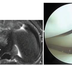
September 24, 2025—According to the American Journal of Roentgenology (AJR), MRI can reliably identify lateral meniscal ...
October 03, 2025 
News | Radiology Imaging
Sept. 10, 2025 — According to ARRS’ American Journal of Roentgenology (AJR), general-purpose large language models ...
September 12, 2025 
News | Prostate Cancer
July 16, 2025 — Artificial intelligence can improve diagnostic consistency and reduce false-positives in prostate cancer ...
July 22, 2025 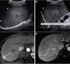
News | Ultrasound Imaging
Jan. 24, 2025 — According to an accepted manuscript published in the American Journal of Roentgenology (AJR), the ACR’s ...
January 28, 2025 
News | PET-CT
July 31, 2024 — In a head-to-head comparison with FDG PET/CT, FDG PET/MRI demonstrated comparable or superior diagnostic ...
July 31, 2024 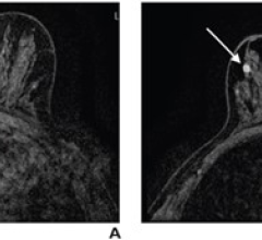
News | Breast Density
June 6, 2024 — Subsequent rounds of abbreviated breast MRI (AB-MR) screening in patients with dense breasts had lower ...
June 06, 2024 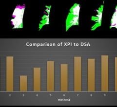
News | Cardiac Imaging
May 17, 2024 — The Cum Laude Award-Winning Online Poster presented during the 124th ARRS Annual Meeting found that the ...
May 17, 2024 © Copyright Wainscot Media. All Rights Reserved.
Subscribe Now