March 27, 2015 — Adding two non-invasive imaging tests to traditional cardiovascular disease risk factor assessment more precisely predicts a healthy patient’s future risk of heart attack, stroke, or premature death, according to a new study. The study was led by Icahn School of Medicine at Mount Sinai and published in the March 24 edition of the Journal of the American College of Cardiology (JACC).
“Using imaging tests to detect disease in carotid or coronary arteries before it causes symptoms can better identify healthy individuals at increased risk than our current, traditional risk assessment methods,” said the study’s principal investigator Valentin Fuster, M.D., Ph.D., director of Mount Sinai Heart and physician-in-chief of The Mount Sinai Hospital.
Study patients were assessed using traditional cardiovascular disease risk factor assessments for high blood pressure, abnormal cholesterol, diabetes, sedentary lifestyle, obesity and smoking; they then additionally received two imaging tests: novel 3-D vascular ultrasound and coronary artery calcium score via a low-dose computed tomography scan. Adding the two imaging tests resulted in the identification of subclinical atherosclerosis in 60 percent of the seemingly healthy study participants presenting with no clinical manifestations.
The BioImage Study was launched to identify new ways to identify subclinical cardiovascular disease before symptoms arise to help curb the growing public health and economic concern of cardiovascular disease. For three years the project followed nearly 6,000 healthy men (ages 55 to 80 years) and women (60 to 88 years) with no history or symptoms of cardiovascular disease.
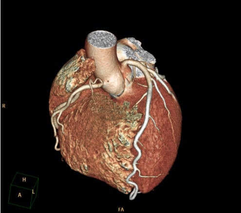
Novel 3-D vascular ultrasound imaging technology allowed researchers to quantify the amount of carotid artery plaque burden lining each patient’s carotid arteries in their neck, while a coronary artery calcium score CT scan allowed for the identification of any narrowing or hardening of the coronary arteries due to the buildup of fatty cholesterol and calcified plaque. Having increased prevalence of carotid plaque burden or coronary artery calcium are signs of atherosclerosis or diseased arteries. Only these imaging procedures could reveal this information in the healthy study participants.
The study shows those apparently healthy individuals identified with increased carotid plaque burden and coronary artery calcium were two to three times more likely to have an adverse event such as an artery blockage, or a piece of plaque becoming loose causing a heart attack or stroke. In fact, after follow-up (2.5 to 3.1 years), there were a total of 216 adverse events reported among study participants including 108 deaths, of which 27 were cardiovascular, plus 34 heart attacks, 30 strokes, 18 experienced unstable angina and hospitalized, and 79 needed revascularization procedures. Over the study duration the incidence of adverse events among those with the greatest amount of carotid and coronary atherosclerosis was 4.2 percent, approximately 8-fold higher compared to individuals with minimal disease in either vascular territory.
“Our study shows the significant impact of adding carotid plaque measurement using vascular ultrasound and coronary calcium scoring with CT scan to our conventional assessment for cardiovascular disease,” said Roxana Mehran, M.D., the study’s co-lead author and director of interventional cardiovascular research and clinical trials at the Zena and Michael A. Weiner Cardiovascular Institute at Mount Sinai Heart at Icahn School of Medicine at Mount Sinai. “Healthy patients with elevated calcium and plaque burden are at increased risk of experiencing adverse cardiovascular events in even a short period of just three years.”
“We must work to identify these patients at risk early and prevent their disease from advancing,” said co-lead author Usman Baber, M.D., assistant professor of medicine, cardiology, at Icahn School of Medicine at Mount Sinai. “Assessing a patient’s risk of atherosclerosis with carotid vascular ultrasound and cardiac calcium CT imaging yields incremental gains over classical risk factors in cardiovascular risk prediction.”
“Using only traditional risk factor assessment for cardiovascular disease may imprecisely classify a patient’s risk,” said Dr. Fuster. “Our study shows simply adding one of our available cardiac imaging resources may more accurately predict a patient’s risk or diagnose their disease, also giving us an opportunity to prevent them from experiencing a future cardiac event and possibly save more lives from the burdens of cardiovascular diseases.”
Fuster added: “For our patients’ lives and for the health of our global economies we need to improve our predictive measures for cardiovascular disease and can no longer just rely on traditional risk factors. Our results prove adding imaging-based biomarkers that directly quantify atherosclerosis are ideal adjuncts to the current conventional CVD risk factors and these imaging biomarkers may be a true game-changer to our practice of cardiovascular medicine.”
According to researchers, their future research will analyze the cost-effectiveness of a new strategy of incorporating these non-invasive imaging tools into future prevention and diagnostic protocols for cardiovascular disease and to better define their use.
For more information: www.icahn.mssm.edu

 June 18, 2026


