Contributing Editor Greg Freiherr offers an overview of computed tomography (CT) advances at the Radiological Society of North America (RSNA) 2015. The video includes Freiherr during his booth tours with some of the key vendors who were featuring new technology.
Technology Report:
Computed Tomography (CT)
Related Content

News | SPECT Imaging
Feb. 5, 2025 — Serac Healthcare Ltd., a clinical radiopharmaceutical company developing an innovative molecular imaging…
 February 05, 2025
News | Computed Tomography (CT)
Computed tomography (CT) has long been a cornerstone of modern imaging, providing detailed 3D insights into the human…
December 19, 2024 
News | Computed Tomography (CT)
Dec. 3, 2024 — During RSNA '24, GE HealthCare announced the 510(k) submission to the U.S. Food and Drug Administration…
December 18, 2024 Sponsored Content
Sponsored Content
Videos | Radiology Imaging
Today’s radiology teams are faced with a wide range of growing complexities such as patient and procedure variabilities ...
June 24, 2026 
News | SPECT Imaging
Dec. 2, 2024 — GE HealthCare has agreed to acquire full ownership of Nihon Medi-Physics Co., Ltd (NMP), by purchasing…
December 05, 2024 
News | Computed Tomography (CT)
Dec. 1, 2024 — Three years after launching its first photon-counting computed tomography (CT) scanner the Naeotom Alpha…
December 02, 2024 
News | Computed Tomography (CT)
Royal Philips recently received 510(k) clearance from the US Food and Drug Administration (FDA) for its detector-based…
November 13, 2024 Sponsored Content

Feature | Information Technology
AT A GLANCE Organization: Expert Radiology Management Services, LLC Specialty: Subspecialty teleradiology — neuro and ...
May 01, 2026 
News | Computed Tomography (CT)
Sept. 9, 2024 — In response to the demand for increased precision in cancer treatments, Beekley Medical has announced…
September 16, 2024 
News | Computed Tomography (CT)
At the annual AHRA (American Healthcare Radiology Administrators) conference in Orlando, Florida, Bayer announced an…
August 09, 2024 
Sponsored Content | News | Computed Tomography (CT)
SPONSORED CONTENT — Fujifilm’s latest CT technology brings exceptional image quality to a compact and user- and patient…
August 06, 2024 Sponsored Content
Sponsored Content
Videos | Radiology Business
Radiology departments have many different needs and face a wide variety of challenges that can impact their departments ...
November 11, 2025 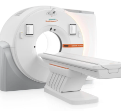
Feature | Computed Tomography (CT) | By Melinda Taschetta-Millane
In the ever-evolving landscape of medical imaging, computed tomography (CT) stands out as a cornerstone technology.…
July 30, 2024 
Videos | Radiology Business
Find actionable insights to achieve sustainability and savings in radiology in this newest of ITN’s “One on One” video…
July 30, 2024 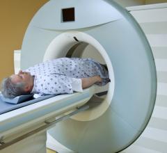
July 24, 2024 — Telix Pharmaceuticals Limited announced that the United States (U.S.) Food and Drug Administration (FDA…
July 24, 2024 Sponsored Content
Feature | Breast Imaging
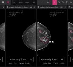
Despite decades of progress in breast imaging, one challenge continues to test even the most skilled radiologists ...
October 24, 2025 
News | RSNA
July 23, 2024 — Professional registration is open for RSNA 2024, the world’s largest radiology forum. This year’s theme…
July 23, 2024 
News | Artificial Intelligence
July 22, 2024 — Healthcare artificial intelligence (AI) systems provider, Qure.ai, has announced its receipt of a Class…
July 22, 2024 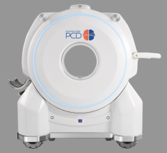
News | Computed Tomography (CT)
July 18, 2024 — NeuroLogica Corp, a subsidiary of Samsung Electronics Co. Ltd., announced its latest configuration of…
July 18, 2024 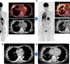
News | PET-CT
July 16, 2024 — A new research paper was published in Oncotarget's Volume 15 on June 20, 2024, titled, “Comparison of…
July 16, 2024 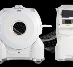
News | Computed Tomography (CT)
July 15, 2024 — NeuroLogica Corp, a subsidiary of Samsung Electronics Co. Ltd., announced its latest configuration of…
July 15, 2024 
News | Prostate Cancer
July 11, 2024 — GE HealthCare’s MIM Software, a global provider of medical imaging analysis and artificial intelligence…
July 11, 2024 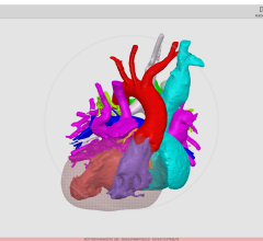
News | Pediatric Imaging
June 25, 2024 — Rady Children’s Hospital-San Diego, one of the nation’s top pediatric health care systems, today…
June 25, 2024 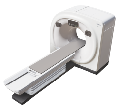
News | Computed Tomography (CT)
June 25, 2024 — Fujifilm Healthcare Americas Corporation, a leading provider of diagnostic and enterprise imaging…
June 25, 2024 © Copyright Wainscot Media. All Rights Reserved.
Subscribe Now