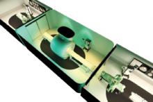
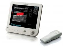
EDAP TMS SA announced the submission of its Pre-Market Approval (PMA) application to the U.S. Food and Drug Administration (FDA) on Jan. 31, 2013 for the Company's Ablatherm-HIFU (high intensity focused ultrasound) for treatment of low risk, localized prostate cancer. EDAP's PMA submission includes data from the ENLIGHT study, a multi-center U.S. Phase II/III clinical trial that completed the two year follow-up needed to evaluate its primary endpoint in August 2012, as well as data from the company's extensive worldwide database of treatment information and follow-up data from patients who have undergone HIFU therapy for prostate cancer.

Dr. John Peixotto of the Carol Milgard Breast Center remembers the old days — before Version 7.0 of the Hologic Physicians Report Writer DX software was installed this past spring — when he was still at the mercy of the dictaphone. Typically, he’d spend an average of three to five minutes per patient dictating bone densitometry test results generated from the Center’s Hologic Discovery system. That may not seem like much. But for a facility like the Milgard Center, which does an average of 20 tests a day, that translated into a lot of extra time crammed into an already busy day. 


Since my first RSNA meeting in 1984, I had been told the only radiologists who had patients were those who did interventions. The mainstream radiologist was a physicians’ physician, providing expert interpretation of medical images, identifying the subtle visual indicators of disease and ruling out diagnoses when signs of pathology were absent. This year was different. 


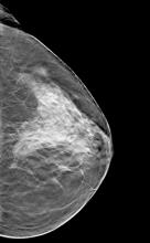
Breast density notification is becoming a reality in many states across the nation by virtue of legislation. A detailed review of breast density is beyond the scope of this article, but there are a few points that bear emphasis.


The Radiological Society of North America (RSNA) annual meeting has transitioned in recent years from an imaging device focus to an imaging information technology focus. The interest in software continued at this year’s meeting, partly fueled by the need for healthcare organizations to meet Stage 1 and 2 meaningful use requirements. Two key trends seen throughout the show floor included remote viewing systems for radiology images and technology streamlined to aid workflow efficiency. 


As other industries turn to the virtual cloud for a variety of information technology (IT) solutions, those in healthcare — especially in radiology — continue to weigh in carefully on the pros and cons of running software tools, as well as hosting their data and medical images, on a third-party server. While the potential for lower costs, improved scalability and faster deployment of services is attractive to physicians and hospitals, concerns such as security issues and increased dependence on an external service provider can also factor into decisions regarding the cloud.


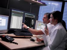
If you would have asked radiologists about the future of radiology information systems (RIS) five to 10 years ago, there is a good chance that they would have told you that RIS was a dying technology. Because RIS systems were merging with other technological platforms, many radiologists did not expect the systems to be around. But now many physicians are trying to meet the Stage 2 meaningful use (MU) criteria to take advantage of Medicare and Medicaid incentive payments and prepare themselves to be in compliance when the criteria become requirements. RIS and picture archiving and communications systems (PACS) are experiencing a technological rebirth that will usher the next generation of these products smoothly into 2013. 


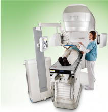
The goal of radiation therapy is to deliver ionizing radiation to eradicate the tumor. Ionizing radiation, however, is indiscriminant and causes damage to both healthy tissues and disease volume along its path. A state-of-the-art treatment would employ intensity-modulated, irregularly shaped beams from various gantry angles to maximize the dose to the target volume and minimize the dose to the surrounding tissues. Accurate beam placement is thus critical to the success of radiation therapy. 


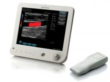
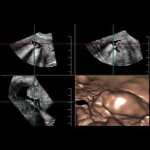
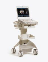
With ever-increasing concerns about radiation dose and the appropriate use of diagnostic imaging tests, doctors are continuously looking for ways to better image their patients. Advances in ultrasound are making the technology appear more attractive for certain clinical applications, from breast health to cardiology, thanks to their noninvasiveness, cost-effectiveness and lack of radiation. Some of the emerging innovations in ultrasound, such as real-time 3-D imaging and the development of wireless transducers, are set to keep the market going throughout 2013 and beyond.


Subscribe Now