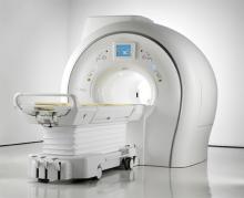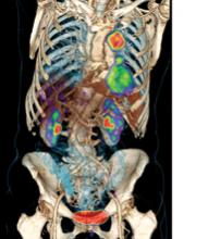
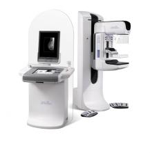
Just weeks after implementing 3D mammography (breast tomosynthesis) in their practice, radiologists at Weinstein Imaging Associates found breast cancers in three patients that were not seen on the 2D images and the entire staff became instant believers in the new technology.  
As a result of routine mammography screening, approximately 1.6 million breast biopsies are performed annually to determine whether a suspicious lesion is malignant or benign. Of these 1.6 million biopsies, approximately 20 percent will result in a positive cancer diagnosis.[1] These numbers are staggering alone, and are just the statistics for one out of more than 200 types of cancers. As radiologists, we strive to provide our patients with the best medical care available, but what we do not always think about is what happens after a sample leaves our office. Errors that occur in the biopsy diagnostic process are specific diagnostic errors that imaging professionals must address. 


Advances in digital mammography are evolving as clinicians look to improve breast cancer screening in women. In recent years, new technological trends have emerged; notably, the addition of 3-D breast imaging, or tomosynthesis, which was approved by the U.S. Food and Drug Administration (FDA) in 2011. But as technology improves, questions remain over the recommended frequency of screening for breast cancer.


For years, high cancer diagnosis rates and limited access to quality health services plagued the Navajo residents cared for by the Tséhootsooí Medical Center in Fort Defiance, Arizona. 


Increasing research on women with dense breasts is having a positive effect on imaging modalities. It is estimated that between 30–40 percent of women in the United States have dense breasts. Because fatty breast tissue makes it difficult to identify breast tumors on standard mammography exams until very advanced stages, dense breasts are a strong independent factor for breast cancer. Research has shown that it is beneficial for this population of women to invest in additional imaging. 


Defining disease processes by their cellular and molecular signatures has enabled molecular diagnosis and the development of targeted therapies, and advances in genomics offer promise for earlier disease detection and possibly molecular prevention. The field of molecular imaging is embracing these advances and is using a diverse set of molecular imaging strategies to interrogate disease states and physiology.


In early February, the Centers for Medicare and Medical Services (CMS) announced a final rule necessary to implement requirements laid out in the Affordable Care Act Sunshine Rule that will increase public disclosure of financial relationships between drug/device manufacturers, and physicians and teaching hospitals.

Subscribe Now