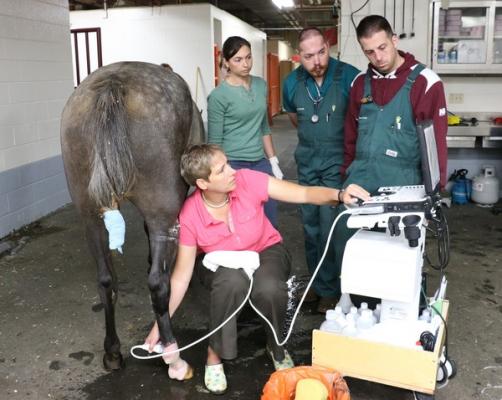
Sabrina Brounts (front), clinical associate professor of large animal surgery, uses ultrasound to gauge how well a Missouri Fox Trotter horse is healing. Photo by Nik Hawkins / Courtesy UW-Madison
September 24, 2014 — An expert in earthquake waves has unearthed the idea that sound waves reflected from human bodies could reveal not only their internal shape but also their condition.
Earthquakes create waves in the earth that are interpreted with seismographs. Medical ultrasound devices make high-frequency waves, and then construct a picture of hidden structures from the echoes. Ray Vanderby, a professor of biomedical engineering and orthopedics and rehabilitation at the University of Wisconsin–Madison, is commercializing an ultrasound method to analyze the condition of soft tissue.
About 10 years ago, Vanderby’s Ph.D. student Hirohito Kobayashi, familiar with seismology and earthquakes, “had an analytical insight into the way waves propagate,” Vanderby said. “I work in orthopedics, so we developed equations that describe the physics of sound waves in living tissue,” he said.
Vanderby has patented the resulting processes through the Wisconsin Alumni Research Foundation, which licensed the technology to Echometrix, a startup that Vanderby and Kobayashi began in 2007. The Echometrix software begins where the ultrasound machine leaves off: by interpreting the image that is the machine’s output. “We can put this software on a laptop and use images from any ultrasound instrument,” said Vanderby.
Conventional ultrasound machines analyze waves that have bounced off a boundary in the body, Vanderby explained. But while ultrasound operators sometimes stretch tissue to get a better picture, the importance of that tactic was not always fully understood. The stretching phenomenon reveals that the intensity of the waves is affected by the condition of the soft tissue. “Waves travel faster in stiff material than in soft material, and the amount of waves that bounce back depends on stiffness, so it was clear that reflected sound was telling us about the stiffness of material,” he said.
In tendons and ligaments, he said, stiffness changes depending on whether the tissue is intact, damaged or healing. “We can measure from the ultrasound image how physically compromised it is,” said Vanderby.
Sabrina Brounts, a clinical associate professor of surgical sciences at the UW–Madison School of Veterinary Medicine, is using the Echometrix technology to study tendon injuries in racehorses.
In 2012, the software was approved by the U.S. Food and Drug Administration (FDA) and is being used in human studies to monitor treatment of injuries to hand tendons, the Achilles tendon and the plantar fascia in the foot. “In plantar fasciitis, it’s hard to tell whether a treatment is helping,” said Vanderby. By producing images that immediately map and quantify damage, he hopes the Echometrix software can measure the pace of healing and identify the best therapies.
But ligaments and tendons — the meat and potatoes of sports injuries — are not the only applications. “We believe any stressed soft tissue could have the same physical behavior,” Vanderby said. “We could use this same technology to detect a change, for example, in tumors or in atherosclerosis, where stiffness indicates degradation of the blood vessel.”
Although magnetic resonance imaging (MRI) equipment can also image soft tissue, “it’s very expensive, it takes longer and the patient has to be constrained,” said Vanderby. “Ultrasound can be used in a dynamic fashion; you can see how it behaves when the tissue is loaded. It’s cheap and it can be done at the point of care.”
The biggest hurdle at Echometrix, Vanderby said, is speeding up the analysis. “The original software was written by a graduate student for research, and we did not care if it took a while. But we’ve made continual progress in accelerating the analysis.”
For more information: www.wisc.edu

 May 07, 2026

 Clearance")


