July 2, 2021 — Philips has participated in an important research project to develop a magnetic resonance (MR) imaging technique [1,2] that could potentially revolutionize the use of MR imaging in cardiology.
Reducing the procedure time for full evaluation of heart anatomy and function from about one hour down to a few minutes, this new technique has the potential to increase patient access to precision diagnosis, improve patient comfort due to shorter scan times, and lower the cost of care. The technique can be used with existing phased-array MRI scanners without modification. The results of a clinical trial to evaluate the technique [2] were published in April, 2021, in JACC (Journal of the American College of Cardiology): Cardiovascular Imaging, one of the world's highest impact journals in the field [3].
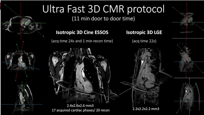
“In just over 20 seconds, all the information needed to know the shape and function of the heart has been acquired. And if you need to evaluate the degree of fibrosis after cardiac muscle death, another 20-second acquisition is all it takes, completing the cardiac study in less than a minute,” said Philips scientist Javier Sánchez-González, Ph.D., technical leader of the Philips team that contributed to the development and leader of the collaboration with CNIC.
During a conventional MR cardiac examination, patients are required to lie still inside the bore of the scanner for about one hour to accurately measure the function of their heart and assess the extent of damaged heart muscle. It requires multiple complex 2D and 3D image acquisitions that need to be captured and reconstructed. As a result, despite being non-invasive and involving no radiation exposure, MR imaging is still not widely used for cardiac imaging.
“The main cause is the time needed to do a full study. A complete study requires about an hour, a period that causes many patients not to finish the test due to the discomfort it causes them,” said Sandra Gómez-Talavera, M.D., researcher at the Spanish National Center for Cardiovascular Research (CNIC), cardiologist at the Hospital Universitario Fundación Jiménez Díaz (Madrid, Spain), and co-author of the JACC paper.
The new technique (called ‘Enhanced SENSE by Static Outer-volume Subtraction (ESSOS)) makes use of the fact that during a breath-hold, everything within the patient’s chest remains static, except their beating heart. After an initial image of the static part (outer volume) has been captured this MRI data is temporarily removed. The MRI signal of the beating heart can now more easily be subtracted from subsequent scan data, allowing up to four times faster acquisition of a 3D image of the heart. This results in a net acceleration factor of up to 32. Once the dynamic information of the beating heart is reconstructed, the static outer volume images are added back to generate a full 3D cardiac image showing heart anatomy and function, and allowing review from different views with good image resolution. If needed, a second contrast-enhanced isotropic 3D single breath-hold scan can reveal the extent of damage to the patient’s heart muscle.
The results of a clinical trial in which more than 100 patients with various cardiac pathologies were examined using both the conventional and the new MR protocol, with the resulting images being evaluated by expert radiologists, demonstrated excellent agreement between heart function measurements made using each technique, as well as excellent agreement in the images to characterize tissue damage to the patient’s heart muscle [4].
“We have shown in a large group of patients that cardiac MR imaging using this new technology obtains the same parameters as the usual technique but reduces the time that a patient has to be inside the machine by more than 90%,” said Borja Ibáñez, M.D., Director of the Clinical Research Department of CNIC, Cardiologist at the University Hospital Fundación Jiménez Díaz, and clinical leader of the work.
The research project to develop this new cardiac MR protocol was financed by the Carlos III Institute of Health, through a FIS technological development project, as well as a Translational Research Grant from the Spanish Society of Cardiology, the European Research Council (ERC), and the Community of Madrid.
While the collaboration between CNIC and Philips is currently at the research stage, the goal is to bring this ultra-fast and easy cardiac MR technique to clinical settings in the near future and support the common vision of benefiting more patients. Already today Philips’ integrated MR solutions offer new levels of ultra-fast and personalized cardiac MR. Examples include the company’s exam-shortening Compressed SENSE technology, ultra-fast early diagnosis of heart failure with SENC and MyoStrain (by Myocardial Solutions), SmartWorkflow end-to-end workflow solution, and helium-free scanners.
For more information: MR Cardiac imaging | Philips Healthcare
References:
[1] ‘Enhanced SENSE by Static Outer-volume Subtraction’ (ESSOS)
[2] Device for research applications only. Not for clinical use.
[4] Gómez-Talavera S, Fernandez-Jimenez R, Fuster V, Nothnagel ND, Kouwenhoven M, Clemence M, García-Lunar I, Gómez-Rubín MC, Navarro F, Pérez-Asenjo B, Fernández-Friera L, Calero MJ, Orejas M, Cabrera JA, Desco M, Pizarro G, Ibáñez B, Sánchez-González J. (2021). Clinical Validation of a 3-Dimensional Ultrafast Cardiac Magnetic Resonance Protocol Including Single Breath-Hold 3-Dimensional Sequences. JACC Cardiovasc Imaging, doi: 10.1016/j.jcmg.2021.02.03

 July 20, 2026


