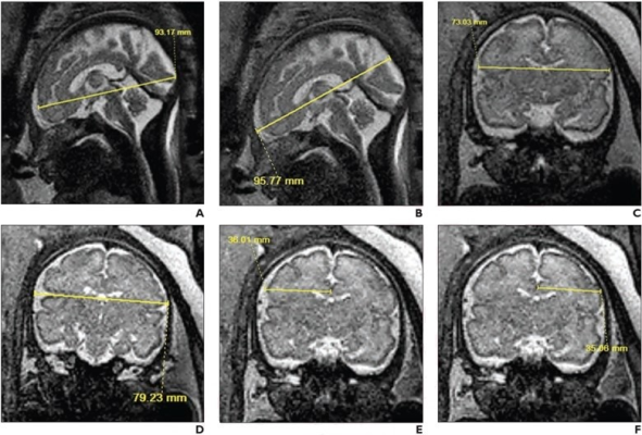
33-year-old patient (gestational age, 30 weeks and 6 days), without in utero opioid exposure, who underwent fetal MRI. Sagittal (A, B) and coronal (C-F) T2-weighted SSFSE images show 2D measurements of brain fronto-occipital diameter (A), bone fronto-occipital diameter (B), brain biparietal diameter (C), bone biparietal diameter (D), right cerebral biparietal diameter (E), left cerebral biparietal diameter (F).
September 29, 2022 — According to an open-access Editor’s Choice article in ARRS’ American Journal of Roentgenology (AJR), third-trimester fetuses with in utero opioid exposure exhibited multiple smaller 2D biometric measurements of the brain, as well as altered fetal physiology, on investigational MRI.
Noting the scarcity of imaging literature evaluating prenatal opioid exposure on brain development, “our findings demonstrate the impact of prenatal opioid exposure on fetal brain development, which may in turn affect postnatal clinical outcomes,” wrote first author Usha D. Nagaraj, MD, from Cincinnati Children’s Hospital Medical Center in Ohio.
Nagaraj and colleagues’ prospective multicenter case-control study included 65 women (mean age, 29 years) in their third trimester of pregnancy who underwent fetal MRI from July 1, 2020 through December 31, 2021 at three U.S. academic medical centers: Cincinnati Children’s Hospital Medical Center, Arkansas Children’s Hospital, and the University of North Carolina at Chapel Hill. A total of 28 fetuses (mean gestational age, 32.3 weeks) were classified as opioid-exposed, while 37 fetuses (mean gestational age, 31.9 weeks) were unexposed in utero. Fourteen 2D biometric measurements of the fetal brain were manually assessed and used to derive four indices.
When adjusting for gestational age, fetal sex, and nicotine exposure, 7 of the 14 2D biometric measurements—cerebral fronto-occipital diameter, bone biparietal diameter, brain biparietal diameter, corpus callosum length, vermis height, anterior-posterior pons measurement, and transverse cerebellar diameter—were significantly smaller in opioid-exposed fetuses than in unexposed fetuses, as measured on fetal MRI.
“In addition,” the authors of this AJR article concluded, “fetuses with prenatal opioid exposure had increased frequencies of breech presentation and of increased amniotic fluid volume.”
For more information: www.arrs.org
Related Fetal MRI Content:
COVID-19 During Pregnancy Doesn’t Harm Baby’s Brain
Researchers Generate 3-D Virtual Reality Models of Unborn Babies


 February 13, 2026


