December 24, 2021 — Newborn babies with heart defects frequently exhibit abnormal extracardiac development of the brain or other organs as well. Fetal MRI, a magnetic resonance imaging procedure that can be performed on the unborn child, can detect these abnormalities at an early stage. In a recent study published in the highly respected Journal of the American College of Cardiology, the team led by Gregor Dovjak from the Department of Biomedical Imaging and Image-guided Therapy in collaboration with the Division of Obstetrics and feto-maternal Medicine at MedUni Vienna and University Hospital Vienna demonstrates the importance of fetal MRIs in the diagnosis of congenital abnormalities.
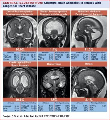
Heart defects are among the most common congenital abnormalities and affect almost 1% of all newborns. Early and comprehensive evaluation of fetuses with sonographically diagnosed heart defects using fetal Magnetic Resonance Imaging (MRI) as well as other techniques is important for the care of the child with a heart defect and the diagnosis of possible further abnormalities, both before and after birth. A new study conducted by the Department of Biomedical Imaging and Image-guided Therapy in collaboration with the Division of Obstetrics and Feto-Maternal Medicine within the Department of Obstetrics and Gynecology of MedUni Vienna and University Hospital Vienna shows that almost 57% of fetuses with heart defects also had at least one other abnormality on the MRI, and approximately a quarter of all fetuses had structural brain abnormalities. According to the study, the severity of the heart defect has no influence on the rate of extracardiac abnormalities in other organs. The study looked at 442 fetuses with heart defects between the 17th and 38th week of gestation.
MRI as a useful complement to ultrasound for the assessment of extracardiac anomalies in fetuses with congenital heart disease
"Currently, fetal MRI is not universally used for prenatal assessment of fetuses with cardiac defects. However, our results suggest that fetal MRI is a useful complement to ultrasound," said Gregor Dovjak, lead author of the study, speaking about the new findings.
Fetal MRI can be used to obtain high-resolution images of the fetal organs, irrespective of the mother's body weight or the amount of amniotic fluid present. This reliable prenatal imaging allows abnormalities to be identified and further treatment steps to be initiated at an early stage. A particular strength of fetal MRI is accurate assessment of structural brain abnormalities that are sometimes difficult to detect using ultrasound. One of the major fetal MRI centres in Europe is located within the Department of Biomedical Imaging and Image-guided Therapy, where several fetal MRI scans are performed on a daily basis.
For more information: www.meduniwien.ac.at/web/
Related Fetal MRI Content:
COVID-19 During Pregnancy Doesn’t Harm Baby’s Brain
Researchers Generate 3-D Virtual Reality Models of Unborn Babies

 July 20, 2026


