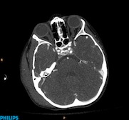
A five-year-old patient fractured the petreous bone after falling at the playground. Ingenuity CT allows low-dose evaluation of the pediatric head and neck with enhanced image quality. Scan obtained at 1.0 mSv.
In emergency and trauma medicine, the “golden hour” refers to the time during which medical/surgical intervention has the greatest likelihood of saving a life. The shorter the time to intervention following a severe injury, the greater the chance of survival. Computed tomography (CT) is integral to these patients’ assessment, diagnosis and treatment planning. Philips Ingenuity CT has advanced features to simplify scanning, reconstruction of images and reporting of results quickly to help improve patient care.
CT is a multi-faceted tool for trauma/emergency, used routinely on new admission of these patients for a multitude of presenting symptoms and histories. There are several types of scans performed on these patients, including: full body, pulmonary CTA, neuro, spine assessment, extremity, abdominal, chest, pediatric, triple rule-out and follow-up scans.
Trauma patients often present with multiple injuries, requiring a full body scan. These patients may have critical injuries and any movement could have dire consequences.Being able to position these patients on the table for a full body scan once without having to further move them for additional scanning is imperative to care.
Many emergency/trauma patients are young in age and could have repeated scans to monitor their progress. It is very important to be able to scan at the lowest possible radiation dose. In addition, scan protocols for emergency/trauma patients often require multi-phase scanning.
Contrast-enhanced exams are complex and are routinely performed on a CT scanner in a trauma/ED setting. A successful CT examination with IV contrast requires synchronization of the scan timing with maximum contrast enhancement in the vessels.
Quick image reconstruction is vital
These CT scans produce a multitude of images and time is of the essence with these patients. Image reconstruction must occur quickly to ensure the radiologist is able to view the images as soon as possible. For correct diagnosis the multiple axial images must be manipulated into sagittal and coronal reconstructions. Automatic production of these images results in consistency and can save the technologist time.
The reliability of the CT scanner is of prime importance in the trauma/ER department because the scanner has to be ready and running to scan patients at any time during the day or night.
Philips Ingenuity CT provides the necessary tools, such as iDose4 iterative reconstruction technique,* and dedicated workflow to perform successful emergency and trauma CT examinations, helping to assure that emergency/trauma CT examinations are performed efficiently and are processed in a timely manner. This can allow the radiologist to provide emergency/trauma physicians and surgeons with diagnoses in the shortest possible time. This can allow the radiologist to provide emergency/trauma physicians and surgeons with diagnoses in the shortest possible time.
Among key features of Philips Ingenuity CT are:
• MRC Ice — The unique spiral grove bearing and slotted anode in the tube allows continuous tube cooling, increasing its life and helping enhance reliability of the scanner. The tube requires no warm up time, so the scanner is always available for scanning patients 24x7 in the ED/trauma department.
• RapidView IR — The new reconstruction engine in Ingenuity CT allows fast reconstruction of images up to 33 images per second, so that images are ready to be reviewed by the physician as soon as the patient is scanned, critical in the trauma/ED setting.
• Dose modulation — The Ingenuity CT scanner has several dose modulation features that reduce the radiation dose delivered to the patient: automatic current setting for each patient based on patient size; longitudinal modulation of the tube current according to patient attenuation at every table position (Z-DOM) and combined longitudinal and angular modulation of tube current (Free-DOM)
• SyncRight — The Philips SyncRight option provides seamless integration between the injector and scanner to synchronize the start of the injector with the scanner.
• In-room controls — The user can position the patient and start/stop the scan after setting up the IV contrast in the scan room. The in-room countdown to X-ray “on” updates the user on the amount of time left to leave the room before the X-ray starts.
• Automatic MPR creation — Results-driven workflow creates automatic MPRs as predefined by the exam card. The user can save the needed MPRs for a particular exam as a part of the exam card for automatic generation after the scan is completed.
• Scannable range — The scanner has a scannable range of 1,850mm, allowing the user to scan from the head to lower extremities without having to move the patient on the table.
• Metal artifact reduction — The metal artifact reduction feature reduces the artifacts generated from metal implants in patient bodies. Patients scanned in trauma/ED may have metal implants and there may not be enough time to collect history and position the patient appropriately. The metal artifact reduction feature reduces artifacts from metal, making it easier to scan these patients.
• Dose reporting — A summary of the radiation dose delivered to the patient is saved with the patient’s exam and sent to PACS for future reference.
• Contrast reporting (only available with SyncRight) — A summary of the patient injection parameters is saved with the patient’s exam and sent to PACS for future reference on the contrast dose delivered to the patient.
• Advanced post-processing — The CT Viewer on the EBW workspace is a single application that allows you to have easy access to axials, create MPRs, volume rendering and other views to simplify your review process.
Case study supplied by Philips Healthcare
* In clinical practice, the use of iDose4 may reduce CT patient dose depending on the clinical task, patient size, anatomical location and clinical practice. A consultation with a radiologist and a physicist should be made to determine the appropriate dose to obtain diagnostic image quality for the particular clinical task.

 June 18, 2026


