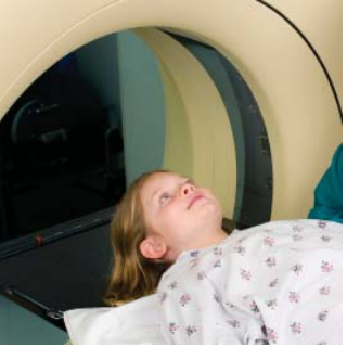
October 4, 2013 — A team of pediatric neurosurgeons and neuroradiologists at the Johns Hopkins Children’s Center has developed a way to minimize dangerous radiation exposure in children with a condition that requires repeat computed tomography (CT) scans of the brain. The experts say they reduced exposure without sacrificing the diagnostic accuracy of the images or compromising treatment decisions.
The approach, described ahead of print in a report in the Journal of Neurosurgery, calls for using fewer X-ray snapshots or “slices” of the brain taken by CT scanners, seven instead of the usual 32 to 40 slices. The approach, the study found, reduced radiation exposure by an average of nearly 92 percent per patient compared with standard head CT scans, while still rendering the images diagnostically accurate.
“The traditional thinking has been that fewer slices would, by definition, mean less clarity and less accuracy, rendering a CT scan suboptimal, but our findings show otherwise,” says lead investigator Jonathan Pindrik, M.D., chief neurosurgery resident at Johns Hopkins.
The research involved analysis of CT scans of patients with excessive fluid in the brain, a condition known as hydrocephalus that requires periodic fluid-draining surgeries and a head CT before each procedure. The investigators compared two standard CT scans with two limited-slice, low-dose CT scans for each one of 50 children, ages 17 and younger, treated for hydrocephalus over five years at Johns Hopkins Children’s Center. The standard CT scan images were performed prior to the launch of the new radiation-minimizing protocol.
In all 50 patients, the radiation-minimizing technique produced clear and 100-percent accurate images of the brain ventricles — the draining system that carries cerebrospinal fluid in and out of the brain. When capturing changes in ventricle size, however, the low-dose approach resulted in a 4 percent error rate: Two of the 50 images were visually ambiguous, generating confusion among the clinicians who reviewed them.
There were no false negatives in the low-dose images, but three false positives, the study showed. In other words, there were no instances of clinicians falsely perceiving normal ventricular size, but three instances of clinicians perceiving a change in ventricular size when there was none. On balance, the radiation-minimizing approach was clearly adequate, the researchers say, and would have not compromised treatment outcomes.
The Hopkins team says the radiation-minimizing technique could be especially valuable in pediatric emergency rooms, where the need for quick diagnosis precludes the use of more cumbersome, radiation-free imaging alternatives like magnetic resonance imaging (MRI). It also can be used for routine evaluation in smaller community hospitals that may lack MRI equipment, the researchers say.
CT scans are invaluable imaging tools that have revolutionized the diagnosis and treatment of many conditions by providing fast, reliable and accurate images, but they have driven up levels of radiation exposure in both children and adults, the investigators say. Ionizing radiation, used in X-rays and CT scans, has been long implicated in the development of certain cancers because of its ability to damage DNA. Children are especially vulnerable to the effects of radiation because of their smaller size, growing tissues and rapidly dividing cells, and because of their longer lifespans that allow slow-growing cancers to emerge decades after exposure, the researchers note.
“We have been searching for ways to minimize radiation exposure in kids without sacrificing the diagnostic accuracy of the images — which is no easy feat — but we believe our limited-slice CT scans achieve that balance,” said senior study investigator Edward Ahn, M.D., a pediatric neurosurgeon at the Johns Hopkins Children’s Center.
Hydrocephalus is often treated by placing a catheter that drains excess fluid from the brain into the abdomen, a procedure known as ventriculo-peritoneal shunting. Children with brain shunts require periodic imaging to assess catheter position and function. Shunt failure that occurs when a catheter dislodges or infection sets in is a common and dangerous complication and considered an emergency. However because shunt failure often causes non-specific symptoms such as headache, vomiting and fever, a definitive diagnosis requires a brain scan.
There are more than 62 million CT scans performed each year in the United States, four million of them in children. Experts estimate that radiation from medical imaging is the single largest source of radiation exposure in the population, and that up to 2 percent of all cancers stem from exposure to medical radiation.
The work was funded by The Hartwell Foundation. Co-investigators included Thierry Huisman, M.D.; Mahadeveappa Mahesh, M.S., Ph.D.; and Aylin Tekes, M.D., all from Johns Hopkins.
For more information: www.newswise.com/institutions/newsroom/63

 June 18, 2026


