January 2, 2018 — Seno Medical Instruments Inc. (Seno Medical) recently reported data from their clinical study demonstrating that its Imagio opto-acoustic (OA) breast imaging system achieved significant differentiation among prognostic subtypes of breast cancer (PSBC). These prognostic subtypes provide important information about the genes that each patient's cancer expresses. The data were presented during a poster presentation at the San Antonio Breast Cancer Symposium, Dec. 5-9 in San Antonio, Texas.
"Confirming a diagnosis of breast cancer is just the first step in understanding the pathology of a patient's tumor," said study presenter Stephen Grobmyer, M.D., director, breast surgical oncology, Cleveland Clinic. "Determining a patient's prognostic subtype of breast cancer is essential for understanding how tumors will respond to specific therapies and selecting a treatment regimen that offers the patient the best chance of a positive outcome. The findings that OA/US [ultrasound] may differentiate among several breast cancer subtypes suggest that this novel imaging methodology could potentially provide a non-invasive alternative to biopsy without sacrificing information about the molecular pathology of the tumor. This could be an important advance for women with breast cancer."
The prospective, multi-center study was conducted at 16 centers in the United States. A total of 1,808 breast masses in 1,739 subjects assessed as BI-RADS (Breast Imaging and Reporting Data System,) category 3, 4 or 5 were imaged with OA/US, resulting in the identification of 678 malignant lesions. Eight blinded readers scored these 678 lesions based on three internal features of the tumor and two external features of the tumor boundary and surrounding tissue. An experienced central breast pathologist who was blinded to the OA/US assessment confirmed the pathologic diagnoses. Biopsy specimens were obtained and assessed for the expression of four genes that comprise the PSBC: estrogen receptor (ER), progesterone receptor (PR), HER2-neu (HER2) and Ki-67. The PSBCs are defined as follows:
- Luminal A (LUM-A): ER/PR (+) and Ki-67 and HER2 (-). These tumors are often low grade, with slow tumor growth and are associated with the most positive prognosis;
- Luminal B (LUM-B): ER (+), PR (+/-), Ki-67 (+) and HER2 (+/-). These tumors are also low grade but grow more quickly than LUM-A;
- Triple-negative breast cancer (TNBC): ER/PR (-), Ki-67 (+/-) and HER2 (-). These tumors are aggressive and typically associated with poor prognosis; and
- HER2-enriched (HER2): ER/PR (-), Ki-67 (+/-) and HER2 (+). These tumors tend to grow quickly but are responsive to anti-HER2 targeted therapies.
Tumors not meeting the definition of any of these categories were deemed unclassified.
Key findings from the study include:
- There was a significant difference in the sum of the three internal OA/US features between LUM-A and TNBC subtypes (p = 0.0256) and borderline significance between LUM-A and LUM-B (p = 0.0831) and LUM-A and HER2 (p = 0.0707);
- There was a significant difference in the sum of the two external OA/US features between LUM-A and TNBC subtypes (p = 0.0001) and LUM-B and TNBC subtypes (p = 0.0020) and borderline significance between LUM-A and HER2 (p = 0.0617);
- TNBC tumors showed mainly internal findings consisting of polymorphic intensely deoxygenated vessels; and
- LUM-A tumors showed a paucity of internal findings and abundant external findings consisting of short perpendicularly oriented tumor vessels in the boundary zone (BZ whiskers), partial BZ deoxygenated blush, and peripheral radiating deoxygenated vessels.
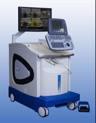
The Imagio OA/US breast imaging system was designed and is being studied to identify two functional hallmarks of cancer: the presence of abnormal blood vessels (tumor angiogenesis) and the relative reduction in oxygen content of blood that occurs in cancer compared to benign masses and normal tissues. The technology is the subject of a U.S. premarket approval (PMA) filing with the U.S. Food and Drug Administration (FDA) and does have European CE Mark.
For more information: www.senomedical.com

 July 15, 2026


