Jan. 27, 2026 — Researchers at the Icahn School of Medicine at Mount Sinai, in collaboration with other leading institutions across the country, have published a new study that provides radiation oncologists with practical guidance to identify and protect female sexual organs during pelvic…
Oncology Diagnostics
News and new technology innovations for oncology diagnostics can be found on this channel.
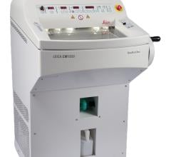
Feb. 11, 2026 — Leica Biosystems has announced the global launch of the Leica CM1950 Cryostat with DualEcoTec Cooling ...
Feb. 3, 2026 — RevealDx, a leader in the characterization of lung nodules, recently announced FDA clearance of RevealAI ...
Jan. 27, 2026 — Researchers at the Icahn School of Medicine at Mount Sinai, in collaboration with other leading ...
For medical practice to advance, efforts to make this practice more efficient must not come at the expense of accuracy ...
Jan. 8, 2026 — RefleXion Medical, an external-beam theranostic oncology company, has announced the U.S. Food and Drug ...
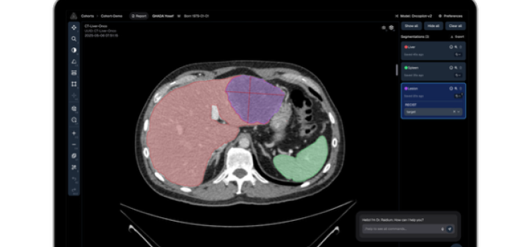
Dec. 1, 2025 — At RSNA 2025, Raidium is introducing its new AI-native PACS Viewer powered by Curia, the first Foundation ...
Nov. 10, 2025 — Researchers at Wayne State University and the Barbara Ann Karmanos Cancer Institute have developed a ...
Efficiency and effectiveness are inseparable in clinical medicine. Digital PET addresses them both. The key is the ...
June 12, 2025 — GE HealthCare has announced the combination of GE HealthCare’s proprietary features and algorithms with ...
Nov. 25, 2024 — CancerIQ has launched a new patient acquisition solution for imaging and oncology, CancerIQ Reach.The ...
Oct. 1, 2024 — At ASTRO 2024, Jay Detsky, MD, PhD, radiation oncologist at Sunnybrook Health Sciences Centre (Toronto ...
Sept. 16, 2024 — IceCure Medical Ltd. has announced the publication of an independent study led by Dr. Franco Orsi ...
May 30, 2024 — Researchers from Penn Medicine’s Abramson Cancer Center (ACC) and the Perelman School of Medicine at the ...
April 2, 2024 — Less than three months after signing an agreement to acquire MIM Software Inc., GE HealthCare ...
February 12, 2024 — Proscia, a leading provider of digital and computational pathology solutions, has received 510(k) ...
December 13, 2023 — The global medical technology company 4DMedical has announced it has entered a binding agreement to ...
September 26, 2023 — Ahead of the 65th American Society for Radiation Oncology Annual Meeting, ASTRO 2023, ITN’s ...
September 14, 2023 — As part of a nationwide action initiative, screening centers nationwide are being asked to open ...
September 7, 2023 — Paige, a provider of end-to-end digital pathology solutions and clinical AI, has announced a ...
August 10, 2023 — Planners and radiation oncology leaders are finalizing programming ahead of the American Society for ...
July 27, 2023 — Privately-held biotech company TAE Life Sciences has announced that its board of directors has appointed ...
March 2, 2023 — Ascelia Pharma, a biotech company focused on improving the life of people living with rare cancer ...




 February 11, 2026


