October 1, 2013 — SuperSonic Imagine’s Aixplorer MultiWave Ultrasound system, first cleared by the U.S. Food and Drug Administration (FDA) in 2009, has received FDA clearance for the quantification capabilities of its real-time ShearWave Elastography (SWE).
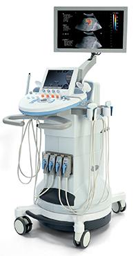
Aixplorer’s UltraFast imaging platform images up to 200 times faster than conventional ultrasound, making it the only system in the world that can, in real-time, generate shear waves in tissue and simultaneously image and compute the velocity of these waves.
Since shear wave velocity is directly related to tissue stiffness, ShearWave Elastography is a technological breakthrough in medical imaging as it permits noninvasive electronic palpation, even in deep, hard to reach organs.
This electronic palpation displays real-time, quantitative (kPa) tissue elasticity on a color-coded map and aids physicians in their diagnostic process, as tissue stiffness varies with the severity of pathology.
ShearWave Elastography imaging also provides additional clinical advantages for ultrasound-guided procedures, evaluation of multifocal stiff tissue, dynamic analysis of elasticity changes and longitudinal follow-up of tissue abnormalities and treatment.
“The clearance of this unique quantification tool for our real-time elastography technology is the result of a close and proactive interaction between the FDA and the company, to substantiate scientific evidence of our product performance,” said Claude Cohen-Bacrie, executive vice president of SuperSonic Imagine. “ShearWave Elastography has been used around the world since 2009 and there are over 50 major publications in several different organs, supporting the clinical benefits of this technique as an adjunct to ultrasound imaging. SWE has been studied extensively in the breast and a large, multicenter clinical trial has demonstrated its added value for lesion characterization.[1] In addition, ShearWave Elastography allows the user to locate stiff nodules in multi-nodule thyroid goiters, to assess the different stages of fibrosis in the liver and to visualize stiff areas in the peripheral zone of the prostate. We are thrilled that American physicians now have access to this invaluable diagnostic tool and we believe the clinical benefits of the Aixplorer will strengthen their medical practices,” he continued.
Wendie Berg, M.D., Ph.D., attending radiologist and investigator stated that, “Being able to actually quantify tissue stiffness in kilopascals, and in a real-time imaging tool, gives clinicians very precise information about the tissue they are scanning. In the case of breast imaging, quantification brings us higher specificity to better evaluate lesions. This translates into better lesion classification that can help avoid some unnecessary biopsies. It can also help us to recognize the need for a biopsy for what was thought to be a probably benign mass.”
Aixplorer’s real-time, quantitative ShearWave Elastography can be seen at SRU in October, AASLD and the RSNA in November 2013.
For more information: www.supersonicimagine.fr
References
1. BE1 clinical study : ShearWave Elastography Improves the Specificity of Breast Ultrasound: The BE1 Multinational Study of 939 masses. Berg WA et al. Radiology 2012. Summary available here.

 May 07, 2026

 Clearance")


