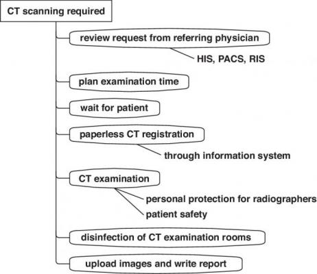
(HIS = hospital information system, RIS = radiology information system) Image courtesy of American Journal of Roentgenology (AJR)
May 18, 2020 — In an open-access article published ahead-of-print by the American Journal of Roentgenology (AJR), a team of Chinese radiologists discussed modifications to the computed tomography (CT) examination process and strict disinfection of examination rooms, while outlining personal protection measures for radiographers during the coronavirus disease (COVID-19) outbreak.
As Jieming Qu, Wenjie Yang, and colleagues at Shanghai Jiao Tong University Medical School Affiliated Ruijin Hospital noted, to undergo CT, patients must exit the fever clinic and proceed to an examination room elsewhere at the institution. Moreover, CT examination rooms are not designed according to the rule of three zones and two aisles — clean zone, semicontaminated zone, contaminated zone; patient aisle and health care worker aisle.
"We were able to urgently install a CT scanner in the fever clinic at the beginning of the outbreak, which allowed rapid screening and early diagnosis," Qu et al. wrote. A safe infection control strategy for examination of patients with suspected SARS-CoV-2 was also implemented, including reconstructing the area and planning the path a patient would take. Additionally, Qu, Yang, and team rerouted the walking pathway to be one-way, limiting ingress and egress while separating contaminated zones from clean zones.
Qu, Yang, and colleagues' extensive routine for examination room disinfection included using an air disinfector (maximum volume of 4000 m3/h) and a movable ultraviolet light (intensity higher than 70 μW/cm2 per meter); cleaning nonplastic equipment surfaces, radiation protection items, and doorknobs with a solution at least 75% alcohol; washing plastic surfaces with soapy solution; and mopping the floor with a disinfectant containing 2000 mg Cl per liter of water. Similarly, all patient waste was considered infectious medical waste and managed accordingly.
Typically, CT scanning is performed by two radiographers. As Qu et al. explained: "The operating radiographer works in the locked control room and controls the scanner (contaminated area). The positioning radiographer works inside and outside the scanning room (contaminated area) and is responsible for communicating with and positioning the patient. The positioning radiographer is not allowed to enter into the control room until the shift ends."
Once a shift is finished, the authors of this AJR article noted that the positioning radiographer is allowed to enter the clean zone only after protective equipment has been properly discarded in the buffer zone.
For more information: www.
Related Coronavirus Content:
VIDEO: Imaging COVID-19 With Point-of-Care Ultrasound (POCUS)
The Cardiac Implications of Novel Coronavirus
CT Provides Best Diagnosis for Novel Coronavirus (COVID-19)
Radiology Lessons for Coronavirus From the SARS and MERS Epidemics
Deployment of Health IT in China’s Fight Against the COVID-19 Epidemic
Emerging Technologies Proving Value in Chinese Coronavirus Fight
Radiologists Describe Coronavirus CT Imaging Features
Coronavirus Update from the FDA
CT Imaging of the 2019 Novel Coronavirus (2019-nCoV) Pneumonia
CT Imaging Features of 2019 Novel Coronavirus (2019-nCoV)
Chest CT Findings of Patients Infected With Novel Coronavirus 2019-nCoV Pneumonia
Find more related clinical content Coronavirus (COVID-19)
ACC COVID-19 recommendations for the cardiovascular care team
VIDEO: What Cardiologists Need to Know about COVID-19 — Interview with Thomas Maddox, M.D.
The Cardiac Implications of Novel Coronavirus
ESC Council on Hypertension Says ACE-I and ARBs Do Not Increase COVID-19 Mortality

 June 24, 2026


