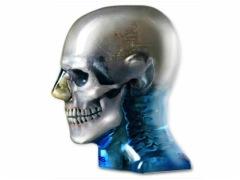
Model 711-HN photo courtesy of CIRS
November 18, 2014 — CIRS will feature a variety of quality assurance (QA), research and training phantoms for all radiology imaging modalities at the 2014 Radiological Society of North America (RSNA) meeting, held Nov. 30-Dec. 3 in Chicago.
Key products that will be showcased include:
UltraiQ Phantom Analysis Software
UltraiQ objectively measures ultrasound image QA parameters and provides faster, more accurate assessments than manual analysis. The software quantitatively and automatically evaluates dynamic range, axial resolution, lateral resolution, caliper distance, dead zone and depth of penetration.
UltraiQ seamlessly integrates with any ultrasound system and can be used with several CIRS Ultrasound Quality Assurance phantoms.
Accreditation Phantoms
Accreditation Uniformity Phantom is a soft, homogeneous mass that conforms to various transducer configurations to evaluate system uniformity.
Accreditation Geometric Accuracy Phantom contains wire targets which facilitate annual assessment of system sensitivity and geometric accuracy as required by the American College of Radiology (ACR).
CBCT Image Quality
Volumetric computed tomography (CT) scanners present unique quality assurance challenges. Noise, resolution, and image uniformity may vary throughout the imaged volume.
With the CIRS Model 062QA-35, each of the four distinct sections is specifically designed with volumetric scanners in mind. This allows comprehensive testing of a full range of image quality parameters.
Dental and Diagnostic Head
The Model 711-HN provides a consistent tool for researchers, clinicians and technologists. It is ideal for determining optimum system settings, commissioning new equipment, monitoring system performance and training in dental X-ray, panoramic X-ray, CT and cone beam CT procedures.
The phantom includes detailed 3-D anthropomorphic anatomy including brain, cortical and trabecular bone, larynx, trachea, sinus, nasal cavities and teeth with dentine, enamel and root structure including the nerve. The sinus cavities are fully open.
For more information: www.cirsinc.com

 October 07, 2026