
March 16, 2022 — The Society of Nuclear Medicine and Molecular Imaging (SNMMI) has named Stanford Health Care and the ...
Molecular Imaging
Nuclear imaging, also called molecular imaging, includes positron emission computed tomography (PET) and single photon emission computed tomography (SPECT) imaging. This section includes radiopharmaceuticals and tracers, PET-CT, SPECT-CT, and PET-MRI. Molecular imaging includes the field of nuclear medicine, which uses very small amounts of radioactive materials, or radiopharmaceuticals, to diagnose and treat disease.

News | Prostate Cancer
March 11, 2022 — NorthStar Medical Radioisotopes, LLC, a global innovator in the development, production and ...
 March 11, 2022
News | Nuclear Imaging
March 4, 2022 — The American College of Radiology (ACR) joins all Americans in calling for an immediate end to fighting ...
March 04, 2022 Sponsored Content

News | PET-CT
Technological advancements in positron emission tomography/computed tomography (PET/CT) offer both clinicians and ...
February 06, 2019 
News | Nuclear Imaging
March 3, 2022 — The Society of Nuclear Medicine and Molecular Imaging (SNMMI) deplores the recent invasion of Ukraine ...
March 03, 2022 
News | Nuclear Imaging
February 18, 2022 — The Nuclear Medicine Europe (NMEu) Emergency Response Team held a call on February 14 on the ...
February 18, 2022 
News | Prostate Cancer
February 18, 2022 — Blue Earth Diagnostics, a Bracco company and recognized leader in the development and ...
February 18, 2022 Sponsored Content

Blog | PET-CT
Efficiency and effectiveness are inseparable in clinical medicine. Digital PET addresses them both. The key is the ...
October 25, 2018 
News | Nuclear Imaging
February 11, 2022 — Due to a recent unexpected reactor shutdown in Europe, the University of Missouri Research Reactor ...
February 11, 2022 
News | Nuclear Imaging
February 2, 2022 — Nuclear Medicine Europe has announced that the HFR reactor in Petten, Netherlands, did not resume ...
February 02, 2022 
News | Molecular Imaging
January 28, 2022 — TeamBest Global Companies (TBG) and Best Cyclotron Systems, Inc. (BCS) announce the shipment of the ...
January 28, 2022 Sponsored Content
Videos | Proton Therapy
Introducing HYPERSCAN , Pencil Beam Scanning Delivery HYPERSCAN enables tumor volumes to be scanned in a matter of ...
October 06, 2014 
News | Nuclear Imaging
January 26, 2022 — On January 24, the Emergency Response Team (ERT) of Nuclear Medicine Europe (NMEu) communicated that ...
January 26, 2022 
News | PET Imaging
January 11, 2022 — Clario, a technology company that delivers the leading endpoint technology solutions for clinical ...
January 11, 2022 
News | Molecular Imaging
Janaury 10, 2022 — The Department of Energy’s National Nuclear Security Administration (NNSA) and Office of ...
January 10, 2022 
News | Molecular Imaging
December 29, 2021 — The global nuclear imaging devices and equipment market is expected to grow from $2.45 billion in ...
December 29, 2021 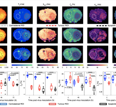
December 27, 2021 — In a recent study published in Nature Biomedical Engineering, a team led by researchers at ...
December 27, 2021 
News | Molecular Imaging
December 7, 2021 — Orviglance* is a novel manganese-based oral contrast agent for magnetic resonance imaging (MRI) ...
December 07, 2021 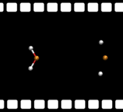
News | Molecular Imaging
December 7, 2021 — An international research team has used the X-ray laser European XFEL to gain new insights into how ...
December 07, 2021 
News | Nuclear Imaging
December 3, 2021 — Mirion Technologies, Inc., a global provider of detection, measurement, analysis and monitoring ...
December 03, 2021 
News | Molecular Imaging
October 29, 2021 — NorthStar Medical Radioisotopes, LLC, a global innovator in the development, production and ...
October 29, 2021 
News | Radiation Therapy
October 19, 2021 — RAD Technology Medical Systems (RAD) announced that it will be exhibiting at the 2021 American ...
October 19, 2021 
News | PET Imaging
October 19, 2021 — Blue Earth Diagnostics, a Bracco company and recognized leader in the development and ...
October 19, 2021 © Copyright Wainscot Media. All Rights Reserved.
Subscribe Now