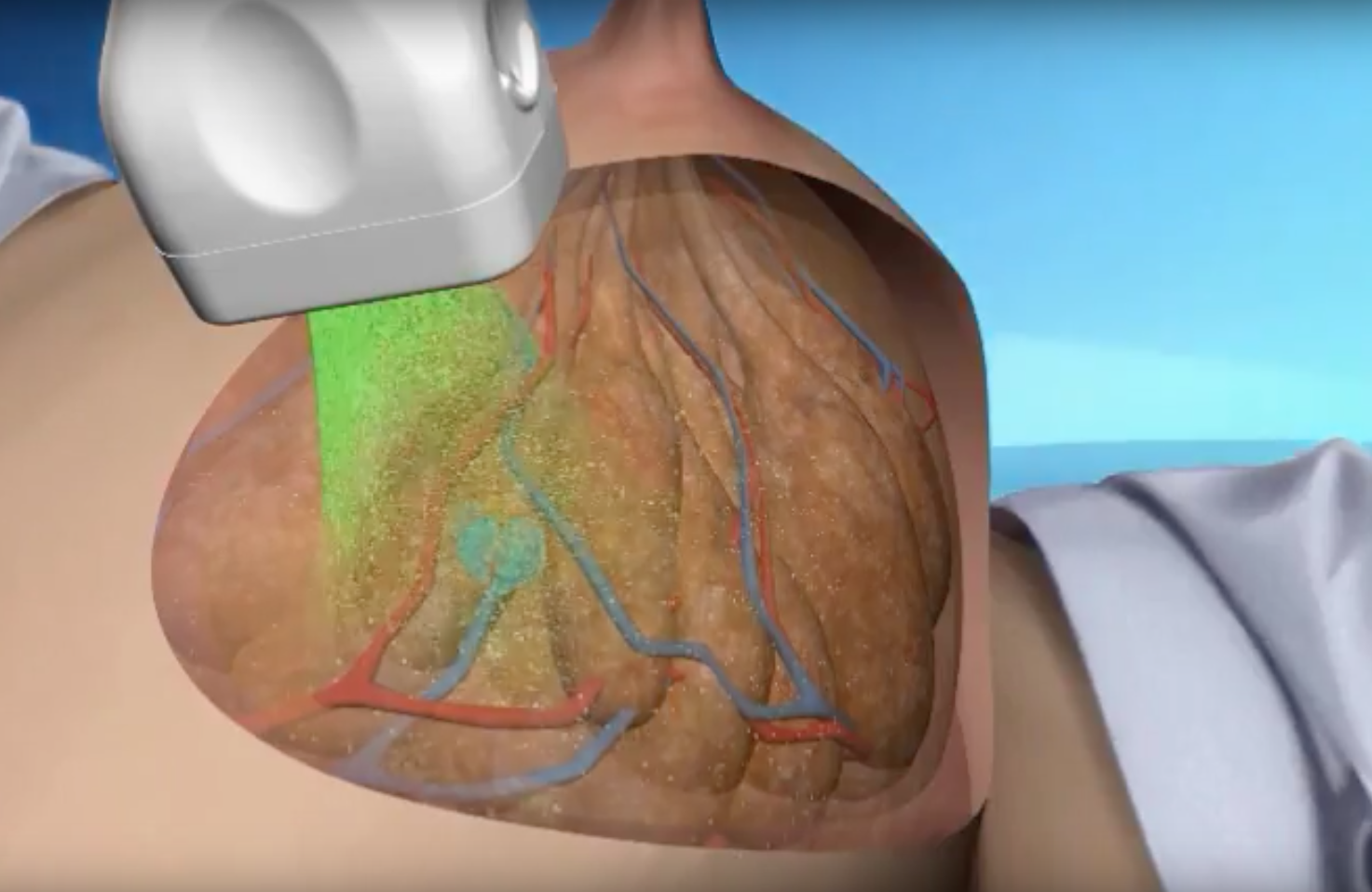
An example of opto-acoustic breast imaging using the Seno system.
January 26, 2017 — There were three high-impact clinical trials presented at the 2017 Radiological Society of North America (RSNA) meeting that offered radiology practice-changing clinical research.
Shared Decision-making in DCIS
For “Decision Quality and Quality of Life After Treatment for DCIS: A Trial of the ECOG-ACRIN Cancer Research Group,” presented by Ruth C. Carlos, M.D., M.S., and Janie M. Lee, M.D., the researchers hypothesized that patient satisfaction with the decision process in cases of ductal carcinoma in situ (DCIS) influences quality of life following treatment. In the study, they assessed quality of treatment decision-making among women with DCIS who were considered candidates for wide local excision and evaluated the impact of decision quality on quality of life after treatment.
The results showed that objective measures of decision quality, such as knowledge score and decision process score, were high. Only knowledge score predicted post-treatment physical and mental well-being. Although there was no significant difference in knowledge score by surgery type, decision process scores were significantly higher, indicating more shared decision-making, for mastectomy compared to wide local excision.
CT Can Better Classify Cardiac Risk
“Core Laboratory Versus Local Site Interpretation of Coronary CT Angiography (CTA): Association with Cardiovascular Events in the PROMISE (PROspective Multicenter Imaging Study for Evaluation of Chest Pain),” presented by Michael T. Lu, M.D., and Jonathan A. Leipsic, M.D., investigates the concordance and relative prognostic value of central core laboratory versus local site interpretation for significant coronary artery disease.
In the PROMISE trial, 193 North American sites interpreted coronary CTA as part of the clinical evaluation of stable chest pain. CTA was also interpreted retrospectively by a central core lab blinded to clinical data, site interpretation, and outcomes. Concordance between core lab and site interpretation for significant CAD (greater than 50 percent luminal stenosis) was assessed.
The findings revealed that core lab interpretation of coronary CTA classified significantly fewer patients as having significant CAD compared to site interpretation, without a loss of predictive power for cardiovascular events.
This study was simultaneously published online in the journal Radiology.
Opto-Acoustic Imaging Better Assesses Breast Masses
The third trial in the session, “A Pivotal Study of Opto-Acoustic Imaging to Diagnose Benign and Malignant Breast Masses: A New Evaluation Tool for Radiologists,” will be presented by Erin I. Neuschler, M.D., and Constance D. Lehman, M.D., Ph.D.
The trial compares the specificity of an investigational optoacoustics (OA/US) breast imaging device with that of gray-scale ultrasound in evaluating breast masses. OA/US functional imaging fuses the high contrast resolution of optical imaging with the good spatial resolution of ultrasound.
A prospective 16-site study was undertaken to compare the BI-RADS classifications of breast masses assigned by independent readers using ultrasound alone to those assigned using OA/US images.
The researchers found that OA/US had better specificity than ultrasound in assessing breast masses and had an excellent negative likelihood ratio. These results suggest that OA/US offers the potential to reduce false positive diagnoses and to further enhance gray scale ultrasound accuracy in breast mass assessment.
This study was published online in the journal Radiology.
Read about other Key RSNA 2017 Study Presentations, Trends and Video.

 July 22, 2026


