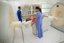
Coupling 50 years of experience with the latest radiological imaging technology and treatments, Princeton Radiology is a global leader in the diagnosis, treatment and research of disease. With five offices located across the state of New Jersey, our practice maintains millions of radiological patient records, including 20 years worth of electronic medical records (EMRs). At Princeton Radiology, we are improving patient care and making data easier to share between offices and physicians. Digital images have changed the way our offices operate.


New studies investigate effectiveness of molecular breast imaging (MBI).


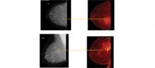
Positron emission mammography (PEM), a high-resolution breast PET scanner, relies on differences in glucose metabolism to identify breast cancers from normal breast cells. Using PEM, we have an opportunity to find cancers at an even earlier stage than that detected with breast MRI, and we may even have the opportunity to find atypia because it changes cellular metabolism prior to the advent of neoangiogenesis. PEM has been shown in recently published prospective data to have similar sensitivity and superior specificity to breast MRI.


Over the years, we have seen an evolution in breast imaging. Screening mammography has been able to detect breast cancer in many women who otherwise have no signs or symptoms. Digital mammography and ultrasound are helping to better diagnose breast cancer in its earliest stages. However, no technology is perfect. Mammography sensitivity is only about 50 percent and decreases to 30 percent in women with dense breasts.
Advanced technologies such as magnetic resonance imaging (MRI) are becoming more widely used for both diagnostic and screening purposes.


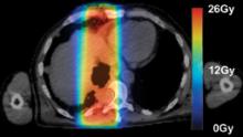
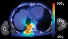
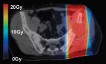
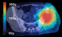
Radiation therapy is an important clinical option for the alleviation of pain and suffering for cancer patients and is used for palliative treatments to prevent pathologic bone fractures or tumor-induced obstructions, bleeding and pain that is resistant to other treatments.

However, standard palliative radiation therapy treatment techniques often offer poor conformality and expose large volumes of normal tissues to radiation-induced toxicities, causing significant side effects for an already ill patient with a limited life expectancy.


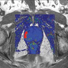
Perhaps one of the most frightening experiences a patient can have is an inconclusive cancer test, where the biopsy is negative, but several other signs point to the possibility that the disease may be present. For some prostate cancer patients, this frightening scenario is a reality.


Treatment planning is a critical component of a patient’s road toward an optimal outcome when dealing with cancer. Whether the ultimate goal is palliative or a total cure, treatment planning always aims to provide just enough dose to a targeted area to eradicate the cancer, while sparing nearby tissue or organs.


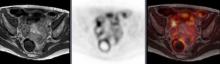
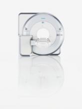
The new imaging modality of positron emission tomography (PET)/magnetic resonance imaging (MRI) was introduced on the U.S, market in June. The Siemens Biograph mMR (molecular MR) gained U.S. Food and Drug Administration (FDA) 510(k) clearance as the first dedicated PET/MRI system. It is also the only system to combine both modalities into one machine, allowing simultaneous imaging of location, function, and metabolic activity of organs in a single image.


June 28, 2011 — Sectra and Philips Healthcare have signed an agreement under which Philips will acquire Sectra’s mammography modality operations for approximately $80 million. Through this deal Sectra’s Medical Systems business will focus on medical imaging information technology (IT).

Subscribe Now