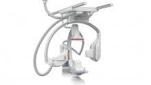
If I lived in a community without 3D mammography, I would strongly advocate my family go to a community offering the 3D exam,” states David Siepmann, M.D., radiologist at Willamette Valley Medical Center. A community hospital located 35 miles south of Portland, Ore., Willamette Valley Medical Center was the first breast center in the state to offer 3D mammography. In the first year, the hospital saw a 21 percent decrease in the overall recall rate and found a number of cancers it might have missed with conventional 2D mammography.


With 2013 on the horizon, ITN looks to see what issues and trends are likely to be prominent for medical imaging in the coming months. Industry experts from several professional societies and organizations gave their input about topics that will be at the forefront in 2013 and their expected impact across major industry segments.


In a comparison of patients with Stage-1 non-small cell lung cancer (NSCLC) during the period 1999-2008, those in the last four years of that period who received radiation therapy had a median survival rate of 21 months, which was markedly better than the 16-month rate among similar patients in the first four years. These study results were presented by lead author Nirav S. Kapadia, M.D., chief resident, department of radiation oncology, University of Michigan Medical School, during the 2012 Chicago Multidisciplinary Symposium in Thoracic Oncology. 


The management of patient information has taken a turn toward complexity with the advent of the new healthcare delivery models as proposed by the Office of the National Coordinator (ONC), meaningful use (MU) and healthcare reform — specifically: Portability and interoperability. The silo and proprietary system model that has served providers for years has been demolished and replaced with a paradigm that requires the sharing of information with affiliated and nonaffiliated providers. The solutions to manage this interoperability are as vast as the HIMSS exhibit hall.


Mobile devices have become a part of everyday life. The cell phone has become as important as your wallet to take with you wherever you go. Some would argue that it’s even more essential now that handheld computers have morphed into a ubiquitous Internet access device for most physicians. 


Imagine being told that complete removal of your left eye was required to stop your spreading cancer. Kathy Kelly of New Milford, Conn., was told just that. An initial biopsy confirmed cancer of her eyelid. In April 2011, surgery revealed Kelly had squamous cell carcinoma, and the disease was not completely removed.


The last decade has seen a significant advancement in imaging technology due to developments in the hardware and software space. It was clear to the radiologists, clinicians and imaging scientists very early on that no single imaging modality, be it magnetic resonance imaging (MRI), computed tomography (CT) or positron emission tomography (PET) could meet all the needs of a clinician treating a patient. 


The introduction of hybrid technology — positron emission tomography/computed tomography (PET/CT) and single-photon emission computed tomography (SPECT)/CT -— has revolutionized the imaging world. This technology allows the combination of the exquisite anatomic details provided, for example, by CT, with the important and much needed functional, physiologic or metabolic information provided by molecular imaging. 


A typical magnetic resonance imaging (MRI) system includes a cryogenic magnet (that creates a static magnetic field), a radiofrequency (RF) coil and resistive electromagnetic gradient coils. The examination time and spatial resolution of MRI are determined by the gradient coils’ speed and strength. 


Computed tomography (CT) vendors have introduced several advancements in hardware and software in 2012. “You want lower dose, better accuracy, and you also want more special and temporal resolution,” said Elliot Fishman, M.D., FSCCT, professor of radiology and oncology, director, diagnostic radiology and body CT, Johns Hopkins University, Baltimore, Md. He said all CT vendors have created next-generation CT scanners to accomplish these goals. New technologies include use of iterative reconstruction, more sensitive detectors that emit less electronic noise, and new software and hardware features to lower dose. New detectors have increased quantum detector efficiency (QDE). The higher QDE, the better the sensitivity of the detector to receive more photons, so lower radiation doses are needed, he explained. 


Subscribe Now