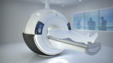
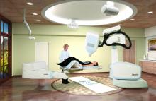
The variety of radiation therapy options continues to grow with the development of treatment planning software, imaging systems for guidance, linear accelerators and more. The external-beam radiation therapy (EBRT) market includes many different types of treatment, from traditional whole-body irradiation (WBI) to conformal radiation therapy (3-D CRT) and intensity-modulated radiation therapy (IMRT). Despite a major decline in 2009, the U.S. radiation therapy market is bouncing back, according to a study published in July by IMV Medical Information Division.[1] Improvements in technologies are helping to drive activity as clinics and hospitals implement advanced techniques to more accurately target and treat cancers.


Beatriz Amendola, M.D., FACR, FASTRO, radiation oncologist, medical director, Innovative Cancer Institute (ICI), decided to work in solo practice in 1999 after moving from Philadelphia to Miami. In Miami, she found there were no privately owned, freestanding facilities offering radiology oncology services to patients. All services were either rendered through a hospital or through a university. That is when Amendola decided to start her own practice, founding the Brachytherapy Center of South Florida in 2002, which is mostly dedicated to the management of early breast cancer and skin cancer using brachytherapy.


In the past 20 years there have been considerable improvements in the treatment of prostate cancer. As it is across all radiation oncology, techniques have evolved from conventional external beam radiation therapy (EBRT), to more targeted therapies that are delivered in fewer visits. One of the latest notable trends in radiotherapy is the emergence of real-time image guidance for intensity-modulated radiation therapy (IMRT). For prostate cancer, radiotherapy is often deployed in conjunction with surgery (prostatectomy) or as curative therapy, depending on the stage of disease. As these new techniques emerge, it becomes more important for clinicians to identify which ones work most effectively and for which patients.


The issue of ionizing radiation in medical imaging is often discussed and is a hotly debated topic. Computed tomography (CT) is the major contributor to medical radiation dose exposure and has been vilified in lay and professional press as a danger to those exposed, potentially carcinogenic and most dangerous in children. As a pediatric radiologist, my primary concern is in producing high-quality diagnostic images with radiation dose as low as reasonably achievable (ALARA).


Injured soldiers returning from combat now have access to more advanced magnetic resonance (MR) technology with Toshiba’s Vantage TitanTM 3T MR. Winn Army Community Hospital in Fort Stewart, Ga., installed the system for general, musculoskeletal and body imaging. This acquisition marks the 1,600th Vantage MR installed worldwide and the 500th installed in the U.S.


Hodgkin lymphoma survivors who received certain radiation and chemotherapy regimens were at increased risk of subsequently developing stomach cancer, according to a study by scientists at the National Cancer Institute (NCI), part of the National Institutes of Health. The study appeared Aug. 26, 2013, in the Journal of Clinical Oncology.


Experts have found that radiation dosage can have serious complications for patients with cancer. Cancer patients who receive chest radiation should be evaluated for heart disease before beginning radiation, and every five to 10 years afterward, according to the American Society of Echocardiography (ASE) and the European Assn. of Cardiovascular Imaging (EACVI) of the European Society of Cardiology (ESC). This expert consensus statement on the use of imaging to detect radiation-induced heart disease (RIHD) will be published in the September issue of the Journal of the American Society of Echocardiography (JASE). The writing group was co-chaired by Vuyisile Nkomo, M.D., an echocardiographer from the renowned Mayo Clinic in Rochester, Minn.


August 28, 2013 — VuComp Inc. announced it has installed its advanced computer-aided detection (CAD) system, M-Vu CAD, at Allison Breast Center in Richmond, Va.  Women in the region will have access to the world’s most advanced tools to help detect breast cancer as early as possible in order to achieve the best outcomes. VuComp’s M-Vu system analyzes mammographic images and marks suspicious areas using sophisticated mathematical algorithms.

August 28, 2013 — Sorna Corp. released the SDS (Secure DICOM Share), a new and innovative feature of the eXpedo software suite. eXpedo with SDS enables healthcare facilities to send DICOM (digital imaging and communications in medicine) studies and reports directly from one site to another through eXpedo, utilizing the DICOM TLS Secure Transport Connection Profile.

Subscribe Now