Photo: Philips
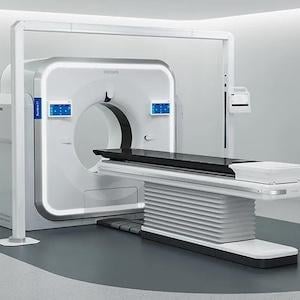
Sept. 17, 2026 — Royal Philips and Southwest Mississippi Regional Medical Center (SMRMC), which serves as the hub of Southwest Health, recently announced that the hospital has installed the first Philips Rembra CT system for acute and high-demand imaging environments in the United States. The system is designed to support the McComb hospital’s high-demand emergency imaging environment with the goal of expanding local access to advanced diagnostic services for patients throughout southwest Mississippi.
SMRMC serves small towns and rural communities across the region and is now using Rembra CT in its emergency and general radiology departments. Designed to accelerate scanning and image reconstruction, Rembra may reduce the time patients spend on the table and help clinicians manage high patient volumes in emergency settings, where every minute matters.
“As healthcare continues to evolve, our goal is to invest in technology that not only advances clinical care but also improves the patient experience,” said Brent Albritton, Director of Radiology at SMRMC. “Our team is already seeing practical day-to-day benefits, including faster system initialization, more efficient image selection, helpful 3D tools and easier patient positioning. These improvements are helping us meet demand while continuing to expand diagnostic services for our community here in McComb.”
Philips Rembra CT
Designed for acute and high-demand imaging environments, the FDA 510(k)-cleared Philips Rembra CT combines advanced detector technology, AI-enabled workflows and rapid image reconstruction designed to make images available in near-real time and support timely clinical decision-making in settings such as emergency and trauma care1. The Rembra CT can support up to 270 patient exams per day in throughput testing, helping imaging departments manage growing patient volumes2.
With the largest-in-class 85 cm bore, the system is designed to help facilitate patient access and positioning [1]. SMRMC also sees an opportunity to further develop its imaging services, including cardiac CT angiography and virtual colonoscopy, as its clinical programs and staffing continue to grow.
“Emergency departments are under constant pressure to move quickly while managing significant patient volumes and clinical complexity,” said Brian Murphy, Vice President, Head of Imaging, North America, Philips. “For community hospitals like SMRMC, access to advanced imaging technology designed for speed, consistency and workflow efficiency can help care teams get the information they need sooner and make more confident decisions when time matters most. With Philips’ Rembra CT, the hospital is strengthening emergency care and helping more patients access advanced imaging close to home.”
References
-
Based on publicly available manufacturer specifications for leading radiology CT systems as of March 2026 on reconstruction speed in this class of scanners and bore size.
-
Based on a 16-hour day and throughput testing of 203 patient scans over 12 hours using typical radiology profiles and protocols.
 October 09, 2026