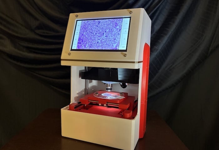
SCANIMUS, the latest iteration of scanning microscopy technology developed by FanousPhotonics, supports both classification and image reconstruction in pathology. (All photos: FanousPhotonics)
As recently as a decade ago, pathology technology was expected to become more expensive, larger and more precise. Yet in the era of artificial intelligence (AI), the opposite seems to be holding true.
“There’s this new trend of innovation, precisely the opposite of what you would expect, which is courting imperfections,” says Michael John Fanous, PhD, founder and CEO of FanousPhotonics. “Because of deep learning, you’re allowed to make certain concessions or compromises, and then compensate for them after the fact.”
Trading Clarity for Speed
Fanous put one such concession, motion blur, to the test in pathology. BlurryScope, a research prototype, scans tissue slides continuously instead of stopping for each exposure. It then applies deep learning to the blurred images. In a peer-reviewed study published in August 2025, BlurryScope classified HER2 status in breast cancer tissue samples with 79.3% accuracy on a four-class scale and 89.7% accuracy on a two-class scale.1
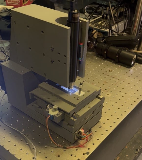
Rather than using a stop-and-stay approach, which halts the slide for each exposure so the image stays sharp, BlurryScope gives up sharpness in exchange for speed. “When we say motion blur, people think it’s some radically different transformation, but it’s usually a very slight difference,” Fanous says.
The same principle — using AI to cull information from incomplete imaging data — is also emerging. Researchers at NYU Langone Health and Meta AI showed that collecting fewer data points and using AI to fill in the rest enabled a twofold reduction in scan time for knee MRIs.2
While image quality is paramount to a radiologist or pathologist, it means something different to an algorithm. “You obviously can’t show a Fourier domain to a pathologist or radiologist,” Fanous says. “That’s just frequency. It’s speckles, and they have no understanding. But to an algorithm, it doesn’t matter. All the algorithm cares about are the numbers.”
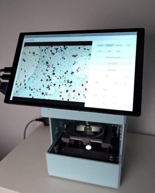
In terms of hardware expenses, BlurryScope, designed solely for immediate classification, costs just $500. Its next-generation successor, SCANIMUS, is a fully integrated unit with onboard processing that supports both classification and image reconstruction. Hardware costs remain relatively modest at $4,500. Initial deployments of SCANIMUS are scheduled for this month.
Addressing Skepticism
Pathologists’ reaction to motion blur, Fanous says, has been “intrigue, followed by a little bit of skepticism.” Overcoming the skepticism involves rigorous training of deep learning models to avoid hallucinations.
“You need to be extremely meticulous because it’s what’s called supervised,” he explains. “I’m sure there’s a lot of this in radiology as well when you’re dealing with reconstructions. You have the labels, the ground truth, and then whatever target compromise or concession there is, and they need to be paired perfectly. If there’s the slightest offset, the whole thing will be off the mark and not usable.”
That level of behind-the-scenes rigor helps turn skeptics into believers. “I think with time, skepticism will start to dissipate because people will realize not only are the reconstructions very good and sharp, but they are going to get better because the model is being trained on systems that are so prohibitively expensive or difficult to operate on,” Fanous says. “In microscopy, we can train the model on super resolution so we can get much better pictures than any scanner.”
Potential Implications for Radiology
While SCANIMUS is solely for pathology, its broader principles could theoretically carry over to diagnostic imaging. “With all of the AI in radiology, you can make scans a lot faster,” Fanous says. The question is, can they be interpreted faster by radiologists? The answer, Fanous says, is “to make sure that the physicians are triaging and facilitating, accelerating the system as much as possible.”
In addition to speed, lower hardware costs could carry over, Fanous says, though each imaging modality (MRI, CT, ultrasound) would need to be tackled differently. “I would say reconfiguring the hardware around the AI, I think that’s the future of it,” he says.
References
-
Fanous MJ, Seybold CM, Chen H, Pillar N, Ozcan A. BlurryScope enables compact, cost-effective scanning microscopy for HER2 scoring using deep learning on blurry images. NPJ Digit Med. 2025;8(1):506. Published Aug. 6, 2025. doi:10.1038/s41746-025-01882-x
-
Johnson PM, Lin DJ, Zbontar J, et al. Deep Learning Reconstruction Enables Prospectively Accelerated Clinical Knee MRI. Radiology. 2023;307(2):e220425. doi:10.1148/radiol.220425

 October 01, 2026