August 7, 2012 — A new method of computed tomography (CT) could produce significantly improved images of knee, spine and hip implants, and may lower radiation exposure, suggested preliminary research presented at the 2012 American Association of Physicists in Medicine (AAPM) annual meeting.
Many of the millions of Americans who receive joint replacements as treatment of arthritis or trauma need CT scans to assess wear, loosening of the prosthesis, fractures or infection, but due to device interference, the images often are tainted by streaks or blurring, which makes diagnosis and assessment of the area around the implant difficult or impossible.
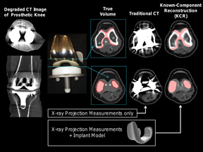
While conventional CT ignores information about the implant, the method developed at Johns Hopkins University in Baltimore, called known component reconstruction (KCR), incorporates a computerized model of the implant’s shape and material content into the 3-D image reconstruction process. Therefore, KCR yields higher image quality and could reduce radiation exposure. Researchers are currently studying the method in clinical CT systems and assessing its potential for routine use in hospitals.
“Every year more than 600,000 people get total knee replacements, which are among the most difficult implants to image around. We truly need a better way to image knee replacements and other implants, and this method is promising,” said co-author J. Webster Stayman, Ph.D., faculty research associate in biomedical engineering at Johns Hopkins University. “This technique is particularly well-suited for implant assessment because surgeons typically know the specific model of the implant. Getting that information into the imaging system could allow them to clearly see tissues around the implant and measure its exact orientation.”
Researchers tested the method in computer simulations and the laboratory using knee implants – one of the most difficult implants to image – as well as surgical screws and rods used in spinal fixation. The results presented at the meeting verified the method using real data and demonstrated that it potentially could be applied generally to CT machines.
“The KCR technique is an exciting advance that combines iterative reconstruction for reduction in radiation dose with strong prior information about implants that are known to be in the image,” said co-author W. Zbijewski, Ph.D., senior research scientist at Johns Hopkins. “We’re working on extending the technique to situations in which the implant changes shape and applying it for the first time to new CT systems for diagnostic radiology and surgery.”
In addition to Stayman and Zbijewski, co-authors of the study presented were Y. Otake, J. Carrino, A. Khanna and J. Siewerdsen.

 June 18, 2026


