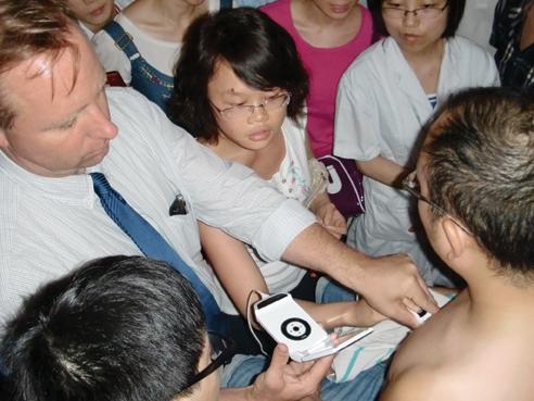
Recently Dr. Thomas Cook taught ultrasound in China, where its usage is in the early stages.
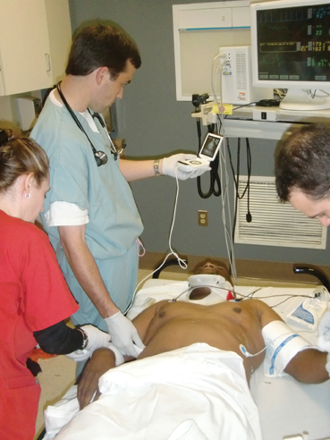
Thomas Cook, M.D., is the emergency medicine residency program director at Palmetto Health Richland in Columbia, S.C., and has been using ultrasound in the practice of emergency medicine since 1996. He also had the recent experience of traveling in China and learning about that country’s use of ultrasound. He shares his expertise and views about ultrasound in the emergency department (ED) in the following Q&A with ITN.
Q: What are the primary uses of ultrasound in the Emergency Department (ED) today?
A: The primary uses for ultrasound in the emergency department are literally anywhere a quick and accurate answer is needed to evaluate and treat our patients. In the 1990s, emergency physicians who were beginning to utilize ultrasound focused on five or six diagnoses, including intraperitoneal fluid from trauma, pericardial effusion, hydronephrosis, gallstones, intrauterine pregnancy and abdominal aortic aneurysm.
Today it’s hard to think of an organ system for which we are not using ultrasound to help with our evaluations. For those areas we were already looking at, the information we glean from examinations has increased enormously. Now, we do not just look for pericardial effusion, but also look at ejection fractions, wall motion abnormalities, valve pathology and the thoracic aorta. Ultrasound in trauma now includes looking for solid organ injury, cardiac function, volume status, pneumothorax, hemothorax and assistance with intravenous access or emergency pericardiocentesis.
Our practice environment requires us to evaluate everything as quickly and efficiently as possible, and there are times when we must do this with limited resources, particularly at night or in rural locations. This creates a situation where the old adage of “necessity is the mother of invention” comes into play. It seems nearly every month someone is publishing an article that describes a new trick to help us manage the myriad of problems we encounter.
Q: What are the benefits of using ultrasound in the ED?
A: It provides what the patients are asking for: speed, accuracy, efficiency, decreased risk of radiation, reassurance and – for lack of better word – it’s cheap. I am always gratified to see how relieved patients are when I can look inside them at the bedside and tell them that everything looks OK. By the same token, when you can quickly find the cause for their visit, it really enhances their impression of your ability to care for them.
From an administrative standpoint, “throughput” is the battle cry for most EDs. As the collective census for the nation’s EDs continues to rise, we are always on the lookout for ways to increase efficiency and get more patients through the system. When you can find the answer at the bedside in minutes rather than using time-consuming (and often less-accurate and more risky) tests, the effect can be dramatic.
Take the case of early pregnancy. It takes no more than one or two minutes to determine that a pregnancy is intrauterine with bedside ultrasound. Now these patients can often be seen in the fast-track.
Q: Is there an issue with ancillary staff training to implement ultrasound use in the ED?
A: From my point of view, it boils down to “why not?” The interpretation of a lot of ultrasound studies is not that difficult.
I am reminded of the early studies with identifying pneumothorax by ultrasound, in which high school students looking at ultrasound exams of the chest had incredible accuracy. In my hospital we have “vascular access teams” that cruise the floors to provide ultrasound-assisted intravenous access in difficult patients. Ask them if they could do their job without ultrasound. (Better yet, ask the patients if they want someone to keep sticking them blindly.)
I am not saying every healthcare worker can use ultrasound for every application, but clearly this technology has proven to be a huge benefit to healthcare workers at many levels. Resistance to using it for a wide array of applications at my hospital is not an issue.
Q: Which types of ultrasound equipment are being used in EDs, and are there changes in how the different types are being used today?
A: This has been and will always be an evolving situation. The only thing you can predict about computers is that they will always be changing. Ultrasound systems in the early ‘90s used to be massive. But with the incredible progress seen in computer miniaturization, we have seen the “point-of-care” ultrasound system become smaller and smaller over a very short span of time.
Is there a tipping point? Is there such a thing as too small? It all depends on what limitations the system imposes due to its size. As I said before, we use ultrasound for anything and everything in the ED. If you make a system that only can accommodate a limited number of studies, emergency physicians will seek out larger systems that offer the full complement of services. These systems will not be the massive, very expensive machines seen in radiology, but will be larger than the hand-held systems currently on the market today.
Most EDs now are using the small laptop or cart-based systems that come with multiple transducers. However, in the long term, the small systems will do everything (much in the way our cell phones are not just cell phones anymore). I predict in the not-too-distant future, ultrasound systems for clinical physicians will become commoditized to the point where the doctor purchases the machine (and everyone has one in his or her pocket).
Q: Where does ultrasound really ‘shine’ in the ED?
A: For me, it’s critical care. As with a lot of emergency departments, the patients are sicker than they have ever been, and you need a quick way to figure out if someone has a life-threatening problem.
I live and work in an area with lots of hypertension, diabetes and renal failure. These patients have lots of vascular disease, and their chief complaints are often “shortness of breath” and “chest pain.” With these patients come the “tough-to-diagnose” problems like pericardial effusion, heart failure versus COPD, pleural effusions, pulmonary edema, volume overload, thoracic aorta pathology and pulmonary embolus. However, I can scan them in a few minutes and get a really good idea whether any of these complaints are present or not.
Another area is vascular access. With all these sick patients, it becomes harder and harder to get adequate intravenous access. Nurses get frustrated, the patient’s care gets delayed and you can’t get the studies you need. We have actually taken it to the level of teaching our nurses to use ultrasound to assist with intravenous access, and this has greatly enhanced our ability to care for these patients quickly.
Q: Where do you see ultrasound use in the emergency department heading in the future?
A: Just about anything you can imagine. There was a time not that long ago where the ultrasound machine was the size of a small refrigerator. I recall telling physicians in the mid-90s that there would come a day when an ultrasound system would be in your pocket and be about the size of a wallet. Of course I sounded nuts at the time, but that day has come and enables possibilities we couldn’t even conjure up a few years ago.
The medical students at the University of South Carolina rotate at our hospital, and each one of them has their own vScan hand-held system for evaluating patients. If you think owning your own system is far-fetched, it’s only the beginning.
For years we have said this technology would be the new stethoscope, but it will be so much more. Beyond the fact that you will not think of going to work without it (regardless of what setting you are seeing patients), it will become your pocket brain as well. It’s only a matter of a few years before this device will integrate with all the hand-held technology we carry around today to help us with patient care.
Also keep in mind that the nation’s medical students over the next decade will be ubiquitously exposed to ultrasound and will integrate its use into how they provide care in the majority of clinical settings. In the future, not using this technology will be like using a typewriter to write a letter.
Q: Beyond the ED — tell us about ultrasound in China
A: I just had the great fortune to travel to China to speak at a meeting on ultrasound use in anesthesia, critical care and emergency medicine. Clinicians there are in the early stages of ultrasound use, but watch out. I was blown away, as most visitors to this country are, but in a country with four times the population of the United States, you must develop technology that is small, cheap and effective if you are going to improve the overall healthcare delivery to your citizens. That ultrasound provides all of this is not lost on them.
So what’s my point? If this huge economic engine adopts ultrasound as quickly it has advanced in every other technology-related field, the use of ultrasound systems, and in particular the smaller hand-held devices, will increase dramatically and likely push ultrasound technology into areas that are not on our collective radar at this time.
Talk to me in a few years and see what I mean. Watch out for the Asian giant!
Thomas Cook, M.D., is currently the emergency medicine residency program director at Palmetto Health Richland in Columbia, S.C., and the founder and educational director of 3rd Rock Ultrasound and the Emergency Ultrasound Course (www.emergencyultrasound.com). He is also a partner in EHEALTHCONCX Healthcare Connectivity Software and Services, Hillside, N.J. (www.portal-us.com), which developed and manages an online review system for ultrasound studies. Cook has been using ultrasound in the practice of emergency medicine since 1996.


 May 07, 2026

 Clearance")


