Getty Images
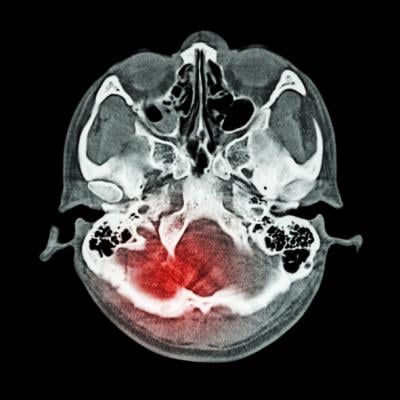
July 21, 2021 — A new study published in JAMA Neurology suggests that certain features that appear on computed tomography (CT) scans help predict outcomes following mild traumatic brain injury (TBI). Patterns detected on the scans may help guide follow up treatment as well as improve recruitment and research study design for head injury clinical trials.
Researchers led by Geoffrey Manley, M.D., Ph.D., professor of neurological surgery at the University of California San Francisco, conducted CT scans in 1,935 subjects with mild TBI and followed their outcomes up to 12 months after injury.
This research was part of the Transforming Research and Clinical Knowledge in Traumatic Brain Injury (TRACK-TBI) study, a large research effort funded by the National Institutes of Health to improve understanding of the short- and long-term effects of head injury and to identify potential treatments.
The researchers identified three distinct sets of patterns on the CT scans, indicating different types of damage after head injury which were associated with various outcomes. The results suggest that contusion (bleeding into brain tissue), subarachnoid hemorrhage (bleeding into the cerebrospinal fluid over the brain), subdural hematoma (bleeding between the brain and the thick covering over the brain), and intraventricular hemorrhage (bleeding into the fluid filled spaces in the center of the brain) were associated with worse outcomes 12 months after injury. Epidural hematoma, which describes bleeding between the skull and outer brain covering known as the dura, was associated with incomplete recovery at two weeks and three months, but was not linked to negative longer-term outcomes.
The TRACK-TBI study was designed and executed in collaboration with the Collaborative European NeuroTrauma Effectiveness Research in Traumatic Brain Injury (CENTER-TBI) study of 2594 TBI subjects. Both sets of results showed similar patterns on CT scans and similar associations between CT abnormalities with clinical outcomes.
More research is needed to understand the effects of head injury on brain structure and function, and how different types of injury can lead to various short- and long-term outcomes.
For more information: www.
Related brain injury content:
MRI Plays Role in Developing Novel Device to Help Protect Athletes’ Brains During Head Impacts

 July 20, 2026


