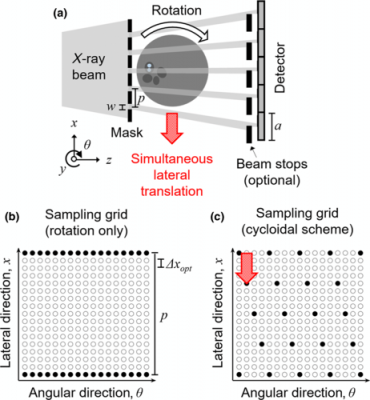
(a) A schematic of cycloidal computed tomography (not to scale, seen from top); by adding an array of beam stops in front of the detector, the setup is transformed into an edge-illumination x-ray phase-contrast imaging device. (b) A sinogram sampling grid for a rotation-only scheme. (c) A sinogram sampling grid for a cycloidal scheme. The grids are shown for one mask period and a subset of rotation angles; the combination of empty and filled circles shows the grids that would be achieved through fine lateral sampling (requiring dithering); the filled circles show the data that are sampled without dithering.
July 24, 2020 — A computed tomography (CT) scan technique that splits a full X-ray beam into thin beamlets can deliver the same quality of image at a much reduced radiation dose, according to a new UCL study.
The technique, demonstrated on a small sample in a micro CT scanner, could potentially be adapted for medical scanners and used to reduce the amount of radiation millions of people are exposed to each year.
A CT scan is a form of X-ray that creates very accurate cross-sectional views of the inside of the body. It is used to guide treatments and diagnose cancers and other diseases.
Past studies have suggested CT scans may cause a small increase in lifelong cancer risk because their high-energy wavelengths can damage DNA. Although cells repair this damage, sometimes these repairs are imperfect, leading to DNA mutations in later years.
In the new study, published in Physical Review Applied, researchers placed a mask with tiny slits over an X-ray beam, breaking up the beam into beamlets. They then moved the sample being imaged in a cycloidal motion that ensured the whole object was irradiated quickly - that is, no parts of it were missed.
The researchers compared the new technique to conventional CT scanning methods, where a sample rotates as a full beam is directed on to it, finding it delivered the same quality of image at a vastly reduced dose.
Charlotte Hagen, Ph.D. (UCL Medical Physics & Biomedical Engineering), first author of the paper and a member of the UCL Advanced X-Ray Imaging Group, said: "Being able to reduce the dose of a CT scan is a long-sought goal. Our technique opens new possibilities for medical research and we believe that it can be adjusted for use in medical scanners, helping to reduce a key source of radiation for people in many countries."
In the NHS, about five million CT scans are performed every year; in the United States, the annual number of CT scans is more than 80 million. CT scanning is thought to account for a quarter of Americans' total exposure to radiation.
Conventional CT scans involve an X-ray beam being rotated around the patient. The new "cycloidal" method combines this rotation with a simultaneous backwards and forwards motion.
The use of beamlets enables a sharper image resolution, as the part of the scanner "reading" the information from the X-ray is able to locate where the information is coming from more precisely.
Professor Sandro Olivo (UCL Medical Physics & Biomedical Engineering), senior author of the paper, said: "This new method fixes two problems. It can be used to reduce the dose, but if deployed at the same dose it can increase the resolution of the image.
"This means that the sharpness of the image can be easily adjusted using masks with different-sized apertures, allowing greater flexibility and freeing the resolution from the constraints of the scanner's hardware."
For more information: www.


 February 09, 2026


