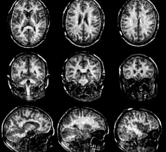
January 3, 2017 — Simon Fraser University researchers have found that high-resolution brain scans, coupled with computational analysis, could play a critical role in helping to detect concussions that conventional scans might miss.
In a study published in PLOS Computational Biology, Vasily Vakorin and Sam Doesburg show how magnetoencephalography (MEG), which maps interactions between regions of the brain, could detect greater levels of neural changes than typical clinical imaging tools such as magnetic resonance imaging (MRI) or computed tomography (CT) scans.
Qualified clinicians typically use those tools, along with other self-reporting measures such as headache or fatigue, to diagnose concussion. They also note that related conditions such as mild traumatic brain injury, often associated with football player collisions, do not appear on conventional scans.
"Changes in communication between brain areas, as detected by MEG, allowed us to detect concussion from individual scans, in situations where MRI or CT failed," said Vakorin. The researchers are scientists with the Behavioral and Cognitive Neuroscience Institute based at SFU, and SFU's ImageTech Lab, a new facility at Surrey Memorial Hospital. Its research-dedicated MEG and MRI scanners make the lab unique in western Canada.
The researchers took MEG scans of 41 men between 20-44 years of age. Half had been diagnosed with concussions within the past three months.
They found that concussions were associated with alterations in the interactions between different brain areas — in other words, there were observable changes in how areas of the brain communicate with one another.
The researchers say MEG offers an unprecedented combination of "excellent temporal and spatial resolution" for reading brain activity to better diagnose concussion where other methods fail.
Relationships between symptom severity and MEG-based classification also show that these methods may provide important measurements of changes in the brain during concussion recovery.
The researchers hope to refine their understanding of specific neural changes associated with concussions to further improve detection, treatment and recovery processes.
The research was funded by Defence Research and Development Canada (DRDC).
For more information: www.journals.plos.org/ploscompbiol


 February 13, 2026


