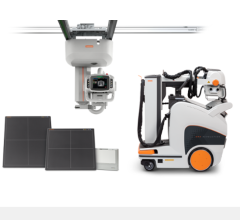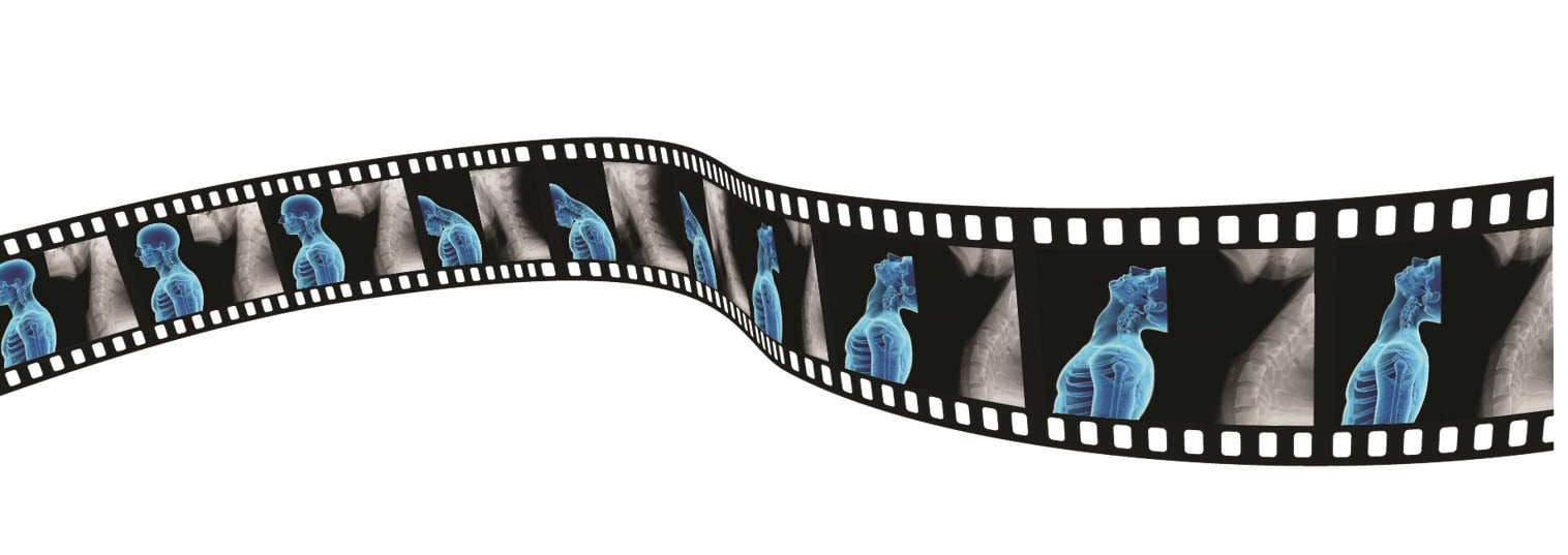
DDR allows clinicians to observe movement like never before. This enhanced version of a standard digital radiographic system can acquire up to 15 sequential radiographs per second resulting in 20 seconds of motion and multiple individual radiographic images. DDR is not fluoroscopy; it is cineradiography, or X-ray that moves. The resulting images provide clinicians with a 4-D data set (a video) that depicts physiological movement.
Musculoskeletal injuries can be difficult to diagnose with a traditional X-ray because X-rays only reveal a static image of a single moment in time. By adding the element of time and motion, Dynamic Digital Radiology (DDR) improves on traditional X-ray by capturing a series of individual digital images at high speed and low dose. The resulting cine loop provides a dynamic look at anatomical structures and a window into a joint in motion.
“You can see how the different parts of the joint itself are moving in relation to each other, and in certain pathologies, some parts don’t move,” said Eric Wagner, M.D., orthopedic surgeon at Emory Healthcare. “A picture is worth a thousand words and a video is worth a thousand pictures. You’re getting more and more critical information from this dynamic motion analysis of
the joint.”
For more than a year, Wagner and a team of orthopedic surgeons at Emory Healthcare have partnered with Konica Minolta to evaluate the use of DDR in patients with musculoskeletal injuries. DDR imaging shows a joint in motion, providing helpful functional and physiological information that can guide diagnosis and treatment pathways for conditions or pathologies where traditional imaging modalities can’t tell a complete story.
“The fact that you’re seeing how the joint itself is moving when a patient is having pain is an inherent advantage,” Wagner said. “Most of the time, the patient is not coming to you because their shoulder hurts at rest; they’re coming to you because their shoulder hurts when they’re trying to move it for certain activities. This imaging allows you to see how the joint is moving during these activities.”
Game-changing Benefits of DDR
Technologies like computed tomography (CT) and magnetic resonance imaging (MRI) can provide high resolution imaging of bony and soft tissue structures, but don’t show the joint’s movement or take images of a patient in a natural upright position. The DDR system can perform all standard X-rays, and images can be taken with the patient standing, seated or on a table.
“In musculoskeletal care, it’s a game changer,” Wagner said. “We are in the infancy of our understanding of exactly what its potential is. I think it’s going to be something that will not only supplant plain X-rays, but probably will take the place of some of the more advanced imaging. You’ll have an idea of what’s going on without needing a more expensive MRI exam or relatively higher radiation CT scan. For injuries like a massive rotator cuff tear, you can see very clearly what’s happening on DDR to make that diagnosis. It’s not something you would need advanced imaging to do.”
In less than a minute, DDR can acquire up to 15 sequential radiographs per second resulting in 20 seconds of motion and multiple single images. The radiation of a typical DDR exam is about equivalent to two standard X-rays.
The Future Potential of DDR
Wagner expects that the wealth of imaging data from the dynamic movement of the joints, such as motion patterns and angle measurements, will foster the creation of artificial intelligence (AI) algorithms that can provide clinical decision support for diagnosis and treatment plans. These algorithms could then determine treatment guidance or identify which patients are better candidates for surgical procedures.
“For surgery, I’m hoping to use it as a way to predict how people will recover after surgeries like shoulder replacements or rotator cuff reconstructions, where there is a certain active motion limitation beforehand. Some of these dynamic parameters viewed on DDR might be able to predict the degree of motion a patient will regain and potentially even help us to better customize our surgical treatments for patients.”
Wagner also sees potential for DDR as a predictive model to diagnose certain pathologies without advanced imaging. For example, DDR can more clearly identify frozen shoulder, which can be difficult to diagnose.
The look inside a joint in motion can also help guide treatment decisions as a patient moves from diagnosis through the care pathway. “We’ve been looking at fracture healing in the hand and wrist,” Wagner said. “If you can’t see the fracture moving when you’re moving your wrist, it’s probably stable enough to discontinue the splint and start advancing your activity.”
Better Diagnosis and Treatment Plans
This new approach to imaging is helping clinicians better understand patient conditions, and better diagnose and treat patients based on how the joints move. A clinician may suspect a particular pathology based on a static image but seeing the joint in motion may change the theory. As clinicians continue to define normal and abnormal joint motion based on DDR imaging, they can better determine diagnosis and treatment plans.
“When I was in my training, my ability to learn a procedure was so much easier when I was watching a video versus when I was reading a textbook and looking at pictures. It’s the same thing comparing DDR to static X-ray,” Wagner said. “It’s so much more helpful, so much more useful when I’m trying to decide what is going on with that patient and how to best treat them.”
DDR is cost-effective, low dose and provides more information on joint motion than is currently available with existing imaging technologies. Wagner expects DDR to become a routine diagnostic tool for musculoskeletal initial workup and care. An X-ray system is more likely to be in an orthopedic practice than advanced imaging. The addition of DDR to standard X-ray can speed up the time to diagnosis for patients.
“I have little doubt that this disruptive technology will eventually change the way we evaluate and manage musculoskeletal injuries,” Wagner said.
For more information: www.xraythatmoves.com


 January 20, 2026