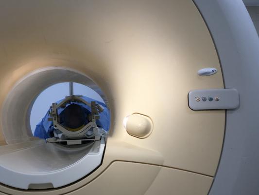
July 5, 2019 — Imaging Biometrics LLC (IB) a subsidiary of IQ-AI Ltd., is highlighting a recently published study in the American Journal of Neuroradiology1 confirming the value of IB’s dT1 maps in streamlining routine radiology workflows and multi-center clinical trials.
The study confirms the value of IB’s dT1 maps in streamlining routine radiology workflows and multi-center clinical trials.
Objective, accurate and reproducible methods to measure brain tumor volumes are important in the assessment of treatment response. Contrast-enhanced magnetic resonance imaging (MRI) is the most common approach to monitor treatment, but it is highly variable and confounded by numerous factors including differences in vendor platforms, field strengths and general MRI system instabilities. It is well-acknowledged that these factors contribute to the large disagreement of up to 50-60 percent between neuroradiologists when assessing tumor burden and evaluating treatment response.
Quantitative dT1 maps offer an elegant and automated solution that overcome these challenges and, therefore, have the potential to cause a disruptive shift in how brain tumor burden is assessed. dT1 maps compare calibrated pre- (T1) and post-contrast (T1+C) anatomic images. The calibration process, exclusive to IB, translates the relative and variable MR intensity values to a fixed and consistent scale. This built-in step is independent of scanner platform, field strength and time point, and has been shown superior over manual tissue normalization algorithms.
The secondary analysis of multicenter clinical trial data was performed by lead author Kathleen Schmainda, Ph.D., from the Medical College of Wisconsin, and colleagues. In total, 123 patients from 23 institutions enrolled in the study. The study compared IB’s dT1 maps to two primary readers and one adjudicator to manually delineate enhancing lesions. The results proved the dT1 method to be comparable to expert reads for determining early tumor progression and proved superior for further distinguishing treatment responders from non-responders at the week 8 time point. Another key outcome of the study was that only the dT1 method could predict differences in outcomes at the week 8 time point.\
For more information: www.imagingbiometrics.com
Reference
1. Schmainda K.M., Prah M.A., Zhang Z., et al. Quantitative Delta T1 (dT1) as a Replacement for Adjudicated Central Reader Analysis of Contrast-Enhancing Tumor Burden: A Subanalysis of the American College of Radiology Imaging Network 6677/Radiation Therapy Oncology Group 0625 Multicenter Brain Tumor Trial. American Journal of Neuroradiology, June 27, 2019. doi: 10.3174/ajnr.A6110

 July 02, 2026


