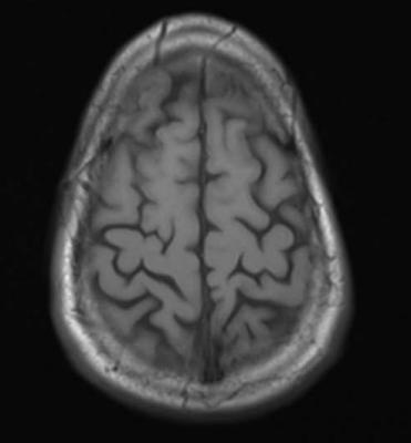
May 19, 2015 — New study results show a simple blood test to measure brain-specific proteins released after a person suffers a traumatic brain injury (TBI) can reliably predict both evidence of TBI on radiographic imaging and injury severity. The potential benefit of adding detection of glial fibrillary acidic protein breakdown products (GFAP-BDP) to clinical screening with computed tomography (CT) and magnetic resonance imaging (MRI) is described in an article published in Journal of Neurotrauma.
Paul McMahon of the University of Pittsburgh Medical Center, and a team of international researchers, including TRACK-TBI investigators, analyzed blood levels of GFAP-BDP from patients ages 16-93 years treated at multiple trauma centers for suspected TBI. They evaluated the ability of the blood-based biomarker to predict intracranial injury as compared to the findings on an admission CT and a delayed MRI scan. The authors reported a net benefit for the use of GFAP-BDP above imaging-based screening alone and a net reduction in unnecessary scans by 12-30 percent in the article "Measurement of the Glial Fibrillary Acidic Protein and Its Breakdown Products GFAP-BDP Biomarker for the Detection of Traumatic Brain Injury Compared to Computed Tomography and Magnetic Resonance Imaging.”
John T. Povlishock, Ph.D., editor-in-chief of Journal of Neurotrauma and professor, Medical College of Virginia Campus of Virginia Commonwealth University, Richmond, noted that "this impressive multi-center study joins with other streams of emerging evidence supporting the use of biomarkers as an important tool in the clinical decision making and prediction process.
"Importantly, this study significantly expands upon other studies that speak to the usefulness of GFAP and, specifically, serum-derived GFAP-BDP in identifying those traumatically brain injured patients whose clinical course is complicated by intracranial injury, demonstrating that GFAP-BDP offers good predictive ability, significant discrimination of injury severity, and net benefit in reducing the need for unnecessary scans, all of which have significant implications for the brain injured patient," said Povlishock.
For more information: www.nationalneurotraumasociety.org

 November 04, 2026