
Sponsored Content | Blog | Digital Radiography (DR) | August 11, 2022
BLOG: Assessing Anatomy in Motion with Dynamic Digital Radiography
A standard X-ray provides tremendous value to a clinician for its ability to quickly provide a static image of anatomical structures. There are limitations, however, to static exams. Multiple conventional X-rays are often needed to show a joint in flexed and extended positions. A single static image cannot provide the details of how a joint moves between flexion and extension points or tell the complete story.
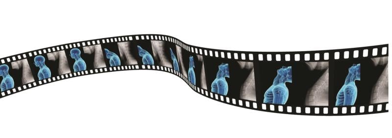
Dynamic digital radiography (DDR) adds dynamic, or motion, capability to X-ray technology that captures sequential radiographic images in a single exam, enabling clinicians to observe the dynamic interaction of anatomical structures in motion over time. Unlike fluoroscopy, X-ray with DDR is not viewed in real-time, so an exam can be performed by a radiologic technologist without a physician present in the exam room.
DDR is an enhanced version of a standard digital radiography system that acquires up to 15 frames per second for as long as 20 seconds, resulting in a maximum of 300 X-ray images with a dose equivalent to about two standard X-rays. With DDR, radiation is lower than fluoroscopy or CT, and requires a shorter exam time than MRI.
Applications in Pulmonology
The benefits of DDR are being explored across a variety of disciplines. In pulmonology, DDR can be used to visualize and quantify lung function in relationship to surrounding structures. “You can watch the patient’s muscles as they breathe – as they inhale and as they exhale,” said Alex Kagen, MD, site chair and associate professor of diagnostic, molecular and interventional radiology at Mount Sinai Morningside and Mount Sinai West in New York City. “You can watch how much air is flowing in and flowing out.”
Dynamic Digital Radiography opens up potential for artificial intelligence and various analytic applications to process the exam, quantify movement and track changes over time. “It’s taking the simple chest x-ray and pairing it with high end technology to now produce a functional exam that wasn’t present previously,” Kagen said.
Applications in Orthopedics
In orthopedics, DDR can be used to assess instability, musculoskeletal injury, sources of pain, and treatment follow-up of any joint throughout its range of motion – whether it be the neck, spine, shoulders or knees. It can also be a helpful tool for postoperative evaluation of movement in place of a more expensive CT or MRI exam.
“The fact that you’re seeing how the joint itself is moving when a patient is having pain is an inherent advantage,” said Eric Wagner, MD, MSc, assistant professor and director of upper extremity research at Emory Healthcare. “Most of the time, the patient is not coming to you because their shoulder hurts at rest; they’re coming to you because their shoulder hurts when they’re trying to move it for certain activities. This imaging allows you to see how the joint is moving during these activities.”
For Wagner, the difference between traditional X-ray and DDR is like learning a procedure from a book versus watching a video tutorial. “It’s so much more helpful, so much more useful when I'm trying to decide what is going on with that patient and how best to treat them,” Wagner said. “I have little doubt that this disruptive technology will eventually change the way we evaluate and manage musculoskeletal injuries.”
The Clinical Value of DDR
DDR use is also being explored as an effective tool for other applications, including swallow studies for speech therapy, or as an alternative to fluoroscopy for assessing diaphragmatic movement during sniff tests.
“Clinicians can now see more with a dynamic X-ray,” said Guillermo Sander, Director of Digital Radiography at Konica Minolta Healthcare. “Because DDR is another X-ray technique using the same X-ray system, the equipment and technician workflow is the same. It just adds an extra minute of time.” DDR is available on the same digital radiography system that performs conventional x-ray exams, providing a cost-effective solution for hospitals and practices.
“For many physicians, DDR provides a more complete diagnosis and has become a key differentiator for their practice,” Sander said. “It introduces a technology with clinical value that also has the benefit of a CPT code for reimbursement.”
In the hospital environment, DDR adds value to what can be done. For patients, there is a wow factor when they see the motion in their X-ray. Sander says that anecdotal feedback suggests that patients who see a dynamic X-ray are more likely to understand their condition and adhere to their rehabilitation plan.
“Adding movement gives new insight, which makes it easier for clinicians to adopt technology and make it useful,” Sander said. “There is clinical value, economic value, technological value and patient appeal. This is a technology worth exploring.”
Editor’s note: This blog is the first in a three-part series about innovative advancements in radiology.
Related DR content:
Guiding Diagnosis and Treatment of Musculoskeletal Conditions With Dynamic X-ray
Related Content

News | Information Technology
July 20, 2026 — GE HealthCare has introduced MIM Anyware, a remote access platform that provides secure, healthcare ...
 July 20, 2026
News | ACR
July 15, 2026 — The American College of Radiology (ACR) recently issued a statement praising the inclusion of the ...
July 16, 2026 
News | Cardiac Imaging
July 8, 2026 — Conavi Medical Corp. has announced the publication of a case report in the Journal of the Society for ...
July 15, 2026 
News | PET Imaging
July 14, 2026 — New research is shedding new light on the biological basis of schizophrenia by directly measuring ...
July 15, 2026 
News | Pediatric Imaging
June 16, 2026 — Crescom has officially launched a global clinical Proof of Concept (PoC) of its pediatric ...
June 24, 2026 
Feature | X-Ray | Kyle Hardner
Water-window X-rays allow researchers to visualize biological cells at high contrast without staining agents or other ...
June 23, 2026 
News | Artificial Intelligence
June 15, 2026 — HOPPR recently announced that HOPPR AI Foundry is now available in AWS Marketplace. The availability ...
June 19, 2026 
News | Radiology Imaging
June 15, 2026 — Lead Glass Pro, a supplier of radiation shielding products, has expanded its turnkey installation ...
June 18, 2026 
News | Digital Pathology
June 15, 2026 — Leica Biosystems is expanding the availability of its Aperio GT Elite digital scanner into the EMEA ...
June 15, 2026 
News | Radiology Business
June 9, 2026 — Bayer has appointed Dr. Jost Reinhard president of the Radiology business within Bayer’s Pharmaceuticals ...
June 12, 2026 © Copyright Wainscot Media. All Rights Reserved.
Subscribe Now