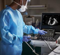
June 23, 2023 — Building on its established leadership in cardiovascular ultrasound, Philips will showcase its ...
Cardiac Imaging
The cardiac imaging channel includes the modalities of computed tomography (CT), cardiac ultrasound (echocardiography), magnetic resonance imaging (MRI), nuclear imaging (PET and SPECT), and angiography.

News | Computed Tomography (CT)
June 22, 2023 — New ultra-high-resolution CT technology enables excellent image quality and accurate diagnosis of ...
 June 22, 2023
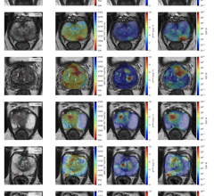
News | Coronavirus (COVID-19)
June 14, 2023 — A University of Waterloo engineer’s MRI invention reveals better than many existing imaging technologies ...
June 14, 2023 Sponsored Content

News | Radiology Imaging
January 23, 2023 — Canon Medical Systems has released a new eBook featuring its new medical imaging roadshow. This new ...
January 23, 2023 
News | Cardiac Imaging
June 9, 2023 — According to an accepted manuscript published in ARRS’ own American Journal of Roentgenology (AJR) ...
June 09, 2023 
June 7, 2023 — GE HealthCare announced the FDA clearance and launch of Sonic DL – a state-of-the-art deep learning-based ...
June 07, 2023 
News | Pediatric Imaging
May 24, 2023 — A new advanced form of computed tomography (CT) imaging called photon-counting computed tomography (PCCT) ...
May 24, 2023 Sponsored Content

Case Study | Cardiac Imaging
In the wake of healthcare reform, facilities have found it essential to offer the most comprehensive solutions to their ...
November 11, 2015 
Feature | Magnetic Resonance Imaging (MRI) | By Johnson Polakkal Joseph
Magnetic resonance imaging (MRI) is a technology that has been around for more than four decades and is a staple in ...
May 01, 2023 
News | Ultrasound Imaging
April 24, 2023 — Butterfly Network, Inc., a digital health company transforming care through the power of handheld ...
April 24, 2023 
News | ARRS
April 18, 2023 — Findings from an award-winning Scientific Online Poster presented during the 2023 ARRS Annual Meeting ...
April 18, 2023 Sponsored Content
Videos | Radiology Imaging
Siemens introduces True volume TEE transducer. This 3-D/4-D 90°x90° TEE solution enables clinically meaningful ...
October 17, 2014 
News | Artificial Intelligence
April 12, 2023 — A global radiology artificial intelligence (AI) company, Annalise.ai, has announced it has received U.S ...
April 12, 2023 
News | ACR
April 4, 2023 — In support of an increased funding recommendation for the National Institutes of Health (NIH) in federal ...
April 04, 2023 
News | PET-CT
March 21, 2023 — Positron Corporation, a molecular imaging device company that offers PET imaging systems and clinical ...
March 21, 2023 
News | Coronavirus (COVID-19)
March 10, 2023 — Researchers found evidence of heart muscle inflammation in a small number of patients with acute ...
March 10, 2023 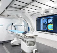
News | SPECT-CT
March 2, 2023 —Royal Philips announced new milestones in the development of the world’s first spectral detector angio CT ...
March 02, 2023 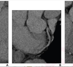
News | Computed Tomography (CT)
March 1, 2023 — According to an accepted manuscript published in ARRS’ American Journal of Roentgenology (AJR), using a ...
March 01, 2023 
February 16, 2023 — According to an accepted manuscript published in ARRS’ American Journal of Roentgenology (AJR) ...
February 16, 2023 
News | Digital Radiography (DR)
February 8, 2023 — Carestream Health is partnering with Robarts Research Institute to increase and demonstrate the ...
February 08, 2023 
News | Computed Tomography (CT)
January 30, 2023 — Photon-counting detector CT reduces the amount of contrast needed for CT angiography (CTA) while ...
January 30, 2023 
Sponsored Content | News | Radiology Imaging
January 23, 2023 — Canon Medical Systems has released a new eBook featuring its new medical imaging roadshow. This new ...
January 23, 2023 
Videos | Cardiac Imaging
Artificial intelligence and general consolidation were two top cardiology trends at RSNA22. ITN/DAIC spoke with Val ...
January 23, 2023 © Copyright Wainscot Media. All Rights Reserved.
Subscribe Now