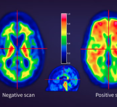
This figure highlights 3 patients with a final diagnosis of low-grade uptake (Perugini grade-1). Endomyocardial biopsy results were available for the patient depicted in Panel B demonstrating ATTR-CA as the underlying pathology. Image created by Department of Nuclear Medicine, Medical University of Vienna, Austria.
January 5, 2023 — A new study has determined that approximately three percent of all bone scan patients have markers of cardiac amyloidosis, which can lead to heart failure and increased mortality. Published in The Journal of Nuclear Medicine, this research is among the first of its kind to identify the prevalence and outcomes of cardiac amyloidosis among the general population.
Cardiac amyloidosis is a disease in which amyloid deposits take the place of normal heart tissue, causing the heart to become thick and stiffen. Formerly believed to be a rare condition, recent diagnostic advances and disease awareness have led to an increasing number of cases of cardiac amyloidosis. Treatment of cardiac amyloidosis is more effective if administered at earlier stages of disease. If left untreated, cardiac amyloidosis can lead to heart failure and death.
“Patients undergoing bone scintigraphy (a nuclear medicine bone scan) may present with high levels of cardiac radiotracer (known as DPD) uptake as an incidental finding, which point to the presence of cardiac amyloidosis,” noted Christian Nitsche, MD, PhD, cardiologist in training and intermediate research fellow at the Medical University of Vienna in Austria and the University College in London, United Kingdom. “In this large-scale study we sought to estimate the prevalence of cardiac amyloidosis in the general population based on bone scans and to investigate the associated outcomes.”
A total of 17,387 scans from 11,527 subjects were analyzed in the study, including both cardiac and non-cardiac referrals. All patients underwent DPD bone scintigraphy, and the scans were analyzed by nuclear medicine professionals. Visual assessments classified the scans as grade-0 (no DPD uptake), grade-1 (low-grade DPD uptake) and grade-2/3 (confirmed cardiac amyloidosis).
Among all subjects, 3.3 percent had some level of DPD uptake (1.8 percent of grade-1 and 1.5 percent of grade-2/3). A prevalence of 1 in 50 among non-cardiac and 1 in 5 among cardiac referrals was reported. There was a significant increase in the prevalence of DPD uptake with incremental age, and comorbidities associated with age (such as arterial hypertension, coronary artery disease and impaired kidney function) were also more prevalent in patients with DPD uptake.
After a median follow-up of six years, nearly 30 percent of subjects died, with cardiovascular death accounting for 8.9 percent of mortality. In addition, 1.5 percent of patients were hospitalized for heart failure during the follow-up period. Patients with grade-2/3 DPD uptake had a 3.5 times higher risk of heart failure hospitalization as compared to those with grade-0 uptake.
“Facing worse outcomes and given the availability of novel treatment options for cardiac amyloidosis, efforts should be maximized to reliably diagnose DPD uptake,” said Andreas Kammerlander, MD, PhD, cardiologist at the Medical University of Vienna in Austria. “Patients with DPD uptake should then be referred to a cardiology specialist for further assessment, as an early diagnosis may offer a window of opportunity to initiate cardiac amyloidosis treatments.
Kammerlander also spoke to the potential for automated detection of cardiac amyloidosis in the future. “Given the advent of novel tracers with high diagnostic accuracy, the diagnosis of cardiac amyloidosis may potentially be established purely non-invasively in the future. With the help of machine-learning algorithms, automated detection of cardiac amyloidosis based on planar imaging alone may represent another possible milestones in the field of molecular imaging.”
For more information: www.snmmi.org


 February 13, 2026


