May 26, 2021 — There are increasing reports of persistent symptoms after a patient recovers from COVID-19, now called long-COVID, lasting for months in some patients. Two of the most common symptoms in patients with long-COVID are fatigue and breathlessness, commonly with no identifiable cause on imaging, lung function or blood tests. In a new study published in Radiology, researchers using a special type of magnetic resonance imaging (MRI) found impaired lung function in patients who had normal chest computed tomography (CT) scans and clinical measurements (D-dimer, hemoglobin and lung function tests) within normal range.[1]
This study by Sheffield and Oxford researchers used a cutting-edge method of imaging has identified persistent damage to the lungs of COVID-19 patients at least three months after they were discharged from hospital, and for some patients even longer. Hyperpolarized xenon-129 MRI (XeMRI) showed abnormalities due to gas transfer limitation in the lungs of patients who were experiencing shortness of breath three months after being discharged from the hospital for COVID-19 pneumonia. XeMRI appears to provide an explanation for patient symptoms not explained by other clinical data or imaging techniques. XeMRI may also provide information on the time it takes for regeneration and recovery to occur.
“The findings of the study are very interesting. The 129Xe MRI is pinpointing the parts of the lung where the physiology of oxygen uptake is impaired due to long standing effects of COVID-19 on the lungs, even though they often look normal on CT scans," explained lead author of the study, Professor Jim Wild, Ph.D., head of imaging and NIHR research professor of magnetic resonance at the University of Sheffield. “It is great to see the imaging technology we have developed rolled out in other clinical centres, working with our collaborators in Oxford on such a timely and clinically important study sets a real precedent for multi-centre research and NHS diagnostic scanning with 129Xe MRI in the U.K.”
“Many COVID-19 patients are still experiencing breathlessness several months after being discharged from hospital, despite their CT scans indicating that their lungs are functioning normally," said study’s principal investigator, Professor Fergus Gleeson. He is a professor of radiology at the University of Oxford and consultant radiologist at Oxford University Hospitals (OUH) NHS Foundation Trust. “Our follow-up scans using hyperpolarised xenon MRI have found that abnormalities not normally visible on regular scans are indeed present, and these abnormalities are preventing oxygen getting into the bloodstream as it should in all parts of the lungs.”
The study, which is supported by the NIHR Oxford Biomedical Research Centre (BRC), has now begun testing patients who were not hospitalised with COVID-19 but who have been attending long COVID clinics.
“Although we are currently only talking about early findings, the XeMRI scans of non-hospitalised patients who are breathless - and 70 per cent of our local patients with Long COVID do experience breathlessness – may have similar abnormalities in their lungs. We need a larger study to identify how common this is and how long it will take to get better.” Professor Gleeson explained.
“We have some way to go before fully comprehending the nature of the lung impairment that follows a COVID-19 infection. But these findings, which are the product of a clinical-academic collaboration between Oxford and Sheffield, are an important step on the path to understanding the biological basis of long COVID and that in turn will help us to develop more effective therapies.”
How Xenon MRI Can Detect Gas Exchange Defects in the Lungs of COVID-longhaulers
Researchers of the study “Hyperpolarized 129-Xenon MRI Abnormalities in Dyspneic Participants Three Months After COVID-19 Pneumonia: Preliminary Results,” undertook this prospective study between August and December 2020, with patients and healthy control volunteers enrolled. All patients underwent lung function tests; ventilation and dissolved phase XeMRI, with the mean red blood cell (RBC):tissue plasma (TP) ratio to be calculated; and a low dose chest CT scored for the degree of post-COVID-19 abnormalities. Healthy controls underwent XeMRI. The intraclass correlation co-efficient was calculated for volunteer and patient scans, to assess repeatability. A Wilcoxon rank-sum test and Cohen's effect size calculated to assess for differences between RBC:TP in patient and controls.
There were 9 patients (mean age 57±7 years, male = 6) and 5 volunteers (29 ± 3 years, Female = 5) enrolled. Patient mean time from hospital discharge was 169, range 116-254 days. There was a difference in RBC:TP between patients and controls (0.3 ± 0.1 versus 0.5 ± 0.1, respectively, p = 0.001, effect size = 1.36).
There was significant difference between the RBC and gas phase spectral full width at half maximum (FWHM) between volunteers and patients (median ± 95 % confidence interval, 567 ± 1 vs. 507 ± 81, p = 0.002 and 104 ± 2 vs. 122 ± 17, p = 0.004, respectively). Results were reproducible with intraclass correlation co-efficients of 0.82 and 0.88 for patients and volunteers respectively. Participants had normal or near normal CT scans, mean 7/25, range 0-10/25.
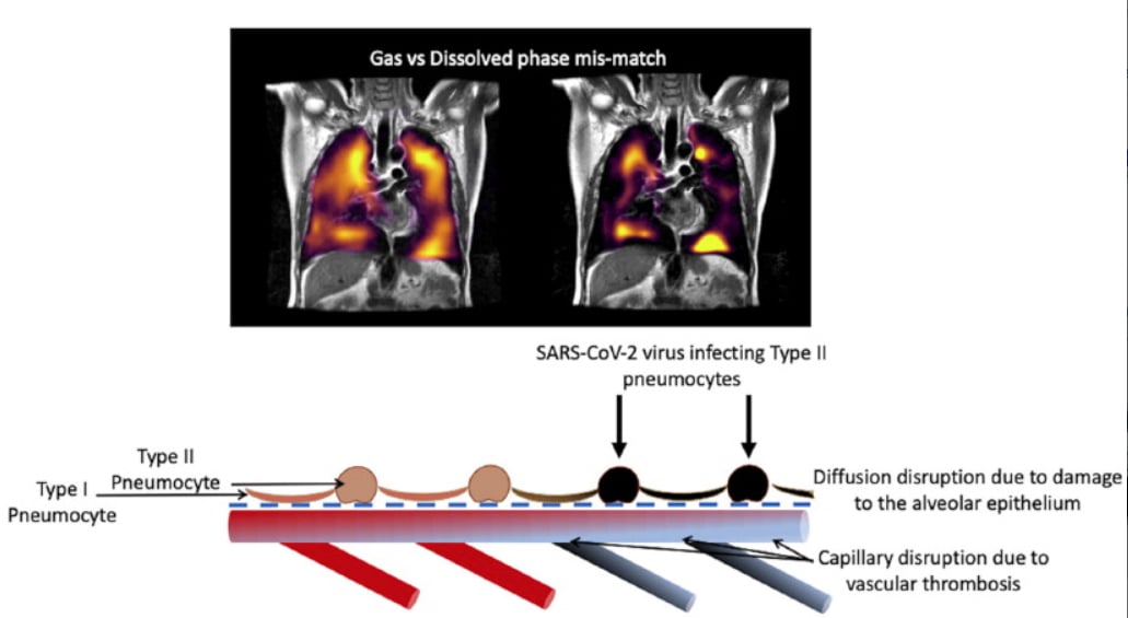
Xe MRI showed alveolar-capillary diffusion limitation in all 9 post COVID-19 pneumonia patients despite normal or nearly normal CT scans, the researchers found.
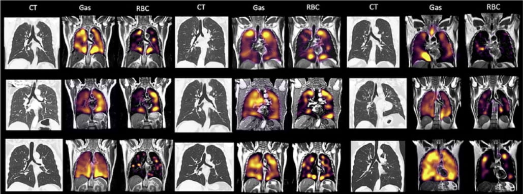
Normal lung CT along with xenon MRI scans showing problems with alveolar-capillary diffusion in 9 COVID-19 long-hauler patients three months after recovering from COVID pneumonia. There is a mismatch in the gas imaging phase (showing concentrations of the xenon gas in the left gas images), and the gas uptake phase where there are numerous areas where there is gas present but it cannot be transferred to the blood due to micro-emboli (seen in the right RBC images). Image courtesy of RSNA.
This Study is Part of the Larger Capturing the MultiORgan Effects of COVID-19 Study in the U.K.
The study, C-MORE-POST in Oxford and MURCO in Sheffield, forms part of the University of Oxford’s C-MORE (Capturing the MultiORgan Effects of COVID-19) study, which feeds into the major national follow-up study PHOSP-COVID, led by the University of Leicester, which is investigating the long-term effects of COVID-19 on hospitalised patients.
The Pulmonary, Lung and Respiratory Imagining Sheffield (POLARIS) research group led by Professor Jim Wild at the University of Sheffield pioneered the methods, development and clinical applications of hyperpolarised gas lung MRI in the U.K., performing the first clinical research studies in the U.K. and the world’s first clinical diagnostic scanning with this technology.
Find more RSNA COVID-19 resources
PHOTO GALLERY: How COVID-19 Appears on Medical Imaging
VIDEO: How to Image COVID-19 and Radiological Presentations of the Virus — Interview with Margarita Revzin, M.D.
Find more radiology related COVID news and video
Reference:

 July 20, 2026


