The ability of magnetic resonance imaging (MRI) to create exquisite images of the body’s soft tissues – and the tumors that arise amid them – is helping physicians at the National Cancer Center Singapore (NCCS) to precisely shape brachytherapy doses to cervical tumors, while at the same time avoiding exposure to critical healthy organs and tissues.
Since November 2011, NCCS clinicians have used MRI to characterize soft tissues, organs at risk and lesions before successive brachytherapy treatments using the microSelectron Digital remote afterloader and Oncentra 3-D brachy image-guided treatment planning system.
Brachytherapy is an advanced, highly targeted cancer treatment in which radiotherapeutic sources are placed in or near a tumor, giving a high radiation dose to the tumor while reducing the radiation exposure in the surrounding healthy tissues. The microSelectron Digital high dose rate (HDR) brachytherapy introduces the radiation source for a certain time using a special applicator.
“After our first year using computed tomography- (CT) based planning for 3-D brachytherapy, we felt confident enough to take the next step: MRI-guided adaptive brachytherapy,” says Richard Yeo, M.D., senior consultant radiation oncologist at NCCS. “The ability to visualize the tumor on MRI and plan the treatment ‘live’ is nothing short of amazing.”
Better Images, Better Treatment
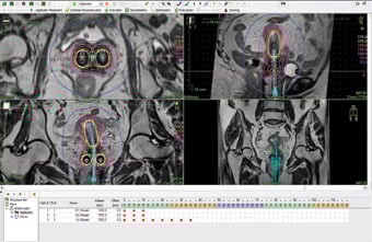
MRI provides information about the tumor’s volume (3-D) and how the volume and shape change between treatment sessions (4-D). Performing an MRI scan before each brachy session enables clinicians to adapt the dose to the unique anatomy of each patient, accounting for not only the position of organs at risk, but also tumor regression or movement, which may have occurred during preceding external beam radiotherapy and/or chemotherapy, and between brachy sessions themselves.
Conforming brachytherapy doses closely to the tumor’s shape and position is important in order to apply the highest possible dose to the tumor while limiting the dose to critical structures, such as the bladder and rectum, thereby decreasing the likelihood of treatment side effects.
Various publications from leading hospitals around the world have shown that use of this advanced technology allows healthcare teams to treat even the most complex cervical cancers, expected to result in lower recurrence rates and higher survival. Intensive multicenter research is ongoing with the aim to provide more data to confirm these treatment benefits over traditional methods. This will allow experts to address unmet medical needs for this common cancer. 1
A group of NCCS experts received training on MRI-guided adaptive therapy at the University Hospital of Vienna, a leading center pioneering the development of this technique for the treatment of cervical cancer. The advanced work done in medical centers in Austria and across Europe is expected to be adopted gradually in a growing number of hospitals in the Asia Pacific region as well, acording to Elekta Brachytherapy,
which manufactures the microSelectron Digital afterloader and Oncentra treatment planning system. itn
1. Radiotherapy and Oncology 94 (2010) 173–180, Int. J. Radiation Oncology Biol. Phys., Vol. 65, No. 2, pp. 624–630, 2006, Clinical Oncology 22 (2010) 602-604.

 July 10, 2026


