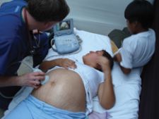
Dr. Tillotson uses the Zonare hand-held z.one ultra system to give a Peruvian woman a first glimpse of her unborn baby.
In the emergency department, time can be a physician's greatest enemy. In a busy setting where many patients present with chest or abdominal pains, there is little to no time to order images and move that patient to another floor for diagnosis.
When time is of the essence and space is tight, versatile hand-carried ultrasound systems are saving lives by providing images in myriad situations – all in a single place, within minutes.
“So often we made decisions with so little information before having hand-held ultrasound,” said Robert Tillotson, DO, RDMS, FACEP, FAAEM, medical director, emergency department, emergency ultrasound director at St. Michael's Hospital, Steven’s Point, WI. “Now I can actually look and see what is going on. I can save the patient unnecessary procedures and when I do procedures, they are considerably safer. Hand-held ultrasound has given me eyes into the body that I have never had before.”
When a trauma patient enters the ED, many questions need quick answers. Is the patient bleeding? Are they bleeding in their abdomen? Is there blood around their heart? Typically, the way to go after these questions would be with needles, invasively, according to Dr. Tillotson, who uses Zonare Medical Systems’ z.one ultra. With a hand-carried ultrasound, a doctor can determine if and where any internal bleeding may be and where to place a central line, laying out the vessel clearly for examination and showing whether or not it is distended or collapsed.
With training, emergency physicians can use hand-carried ultrasounds to quickly image problem areas of the body, instead of waiting for imaging orders to go through.
“The place where [hand-carried ultrasound] has had the biggest impact in my practice is when pregnant patients present with vaginal bleeding,” said Dr. Tillotson. “Before, they would come in, I'd examine them, do a pelvic exam, order an ultrasound, send them down, wait two hours and then get my answer. Now a woman can come into the ER, I can do my exam and ultrasound and if everything looks okay, she goes home in 15 minutes instead of becoming hypertensive in the ultrasound suite.”
The wireless functionality of hand-carried ultrasounds also enables physicians to consult at the patient's bedside so that they can be more informed and involved in their healthcare decisions.
Ultrasounds as standard procedure
The ease of use and fast results of hand-carried devices lend themselves to becoming more common and depended on in emergency
settings. Eyal Herzog, M.D., FACC, director of the Cardiac Care Unit at St. Luke’s-Roosevelt Hospital Center in New York, said all academic, high-level critical care units should now have portable ultrasounds.
“[Hand-carried ultrasound] is very routine now,” said Dr. Tillotson. “I use [the z.one ultra] with pretty much every patient that I see with abdominal pain. I look because I can.”
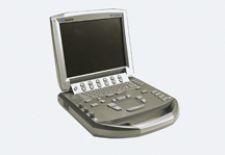
Zonare’s z.one ultra instantly converts from a small, full-featured, cart-based ultrasound system to a compact hand-carried unit without compromising performance. The lap-top sized, 5.5-pound portable system functions on its own or connected to a portable cart system with a large screen.
“The z.one ultra has been phenomenal,” said Dr. Tillotson. “It’s a fantastic machine that gives great images and is quick and easy to use. It has so changed my practice that I approached the state chapter of the American College of Emergency Physicians to propose an introductory course in [hand-carried ultrasound]. I am fired up to help change other practices as well.”
In the ED, there is little to no time to carry a patient to another floor to diagnose a potential heart attack. To address the urgency of diagnosing cardiac conditions, GE Healthcare’s Vivid i is a miniaturized cardiovascular ultrasound system that provides high-performance, full-featured imaging in a lightweight, 10-pound laptop design.
“The Vivid i allows us to do everything in one place,” said Jonathan M. Chen, M.D., director of pediatric cardiac surgery at the Cornell Campus, New York Presbyterian Hospital. “You can take it from room to room, which is huge, and the image is so good.”
Dr. Tillotson said hand-carried ultrasound systems are also being used in nonclinical settings, such as professional hockey, football and the Olympic games for quick diagnostic imaging of sports injuries.
TEE gets to the heart of emergency cardiac imaging
Dr. Herzog routinely uses the Siemens Cypress in his cardiac care unit. “We image almost every patient who comes into our unit,” he said. “If you are sick, it is one of our traditions to first take a look at your heart to get information.”
These hand-carried ultrasound systems also offer connectors for a transesophageal echocardiography (TEE) probe attachment. With TEE, an echo transducer is placed in the esophagus, imaging the heart from behind as opposed to through the ribs. This positioning produces clearer images of the heart, particularly structures such as the left atrium, which may not be seen as well with a transthoracic image.
TEE can be used to detect heart problems such as blood clots, aneurysms, valve dysfunction, septal defects and more.
“We have tested TEE on the Cypress and now use it regularly in our critical care unit,” said Dr. Herzog. “The beauty of this is that I just have to call the echo lab to get a probe and the machine is already in my unit.”
He said using TEE on a portable system is ideal in emergency settings when there is no time to get the patient to a full-sized echo machine.
Mission: possible
The wireless portability and compact size of hand-carried ultrasounds such as the z.one ultra and Vivid i allow clinicians to take the systems out into the field to care for the neediest critical care patients.
Last year, Dr. Tillotson brought the z.one ultra with him on a clinical mission to the impoverished village of Condevilla in Peru. His team examined 1,210 patients in three days. Dr. Tillotson saw a variety of cases, from breech pregnancies to pneumonia, all of which were diagnosed by the Zonare z.one ultra.
“I insisted on taking the z.one with me, because without it I would have been handicapped,” he said. “Because it was battery operated I could use it to diagnose tendon injuries, uterine fibroids, gall stones, placenta previa and all sorts of other problems.”
When Dr. Chen traveled to Senegal with the humanitarian group Surgeons of Hope to help children with congenital heart disease, he depended on GE Healthcare's Vivid i to perform complex operations.
“I don't know if we could have done the trip without [Vivid i],” he said. “If you can't make a correct diagnosis, you cannot perform these sort of procedures. We operated on 10 or 11 kids and the Vivid i allowed us to effortlessly go back and forth between procedures and rooms. A large machine would have been a huge challenge.”
The stethoscope of the future
As the curve of miniaturization technology continues and ultrasound systems become smaller, they may replace common diagnostic tools, such as the stethoscope in both clinical and acute care settings.
“The advantage to the [Siemens Medical Acuson] P10 is that there is always a need to bridge the gap between a traditional stethoscope and a full-sized echo machine,” said Dr. Herzog, who is currently testing the system. “The P10 is filling that gap because you can carry it in your pocket. It is mobile and
easy to use.”

 May 07, 2026

 Clearance")


