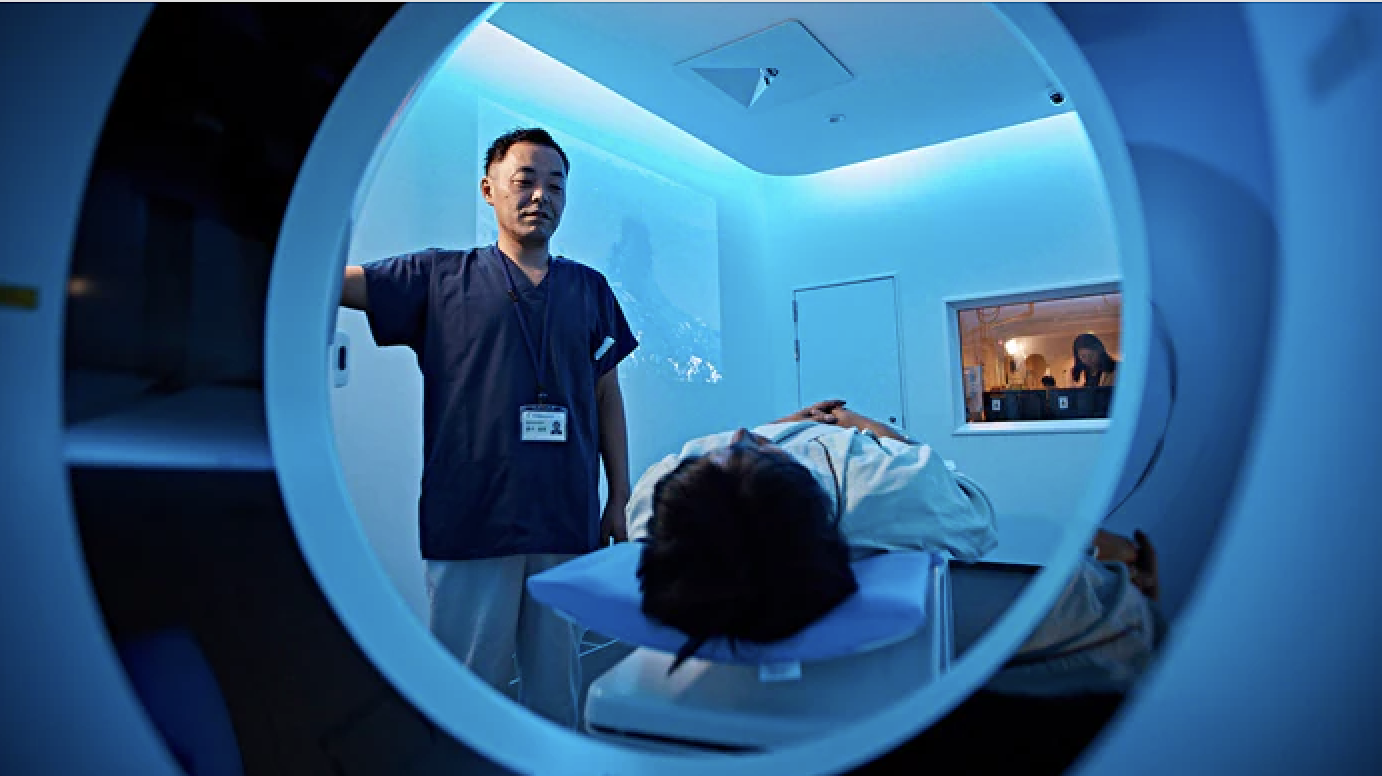
Computed tomography (CT) scans are widely used to diagnose and analyze patients using specialized equipment to generate a sequence of internal body images. The increasing prevalence of chronic diseases and the latest advancements in the healthcare industry is expected to fuel the demand for advanced imaging systems, including CT systems. View the CT Systems Comparison Chart
Mordor Intelligence forecasts that by 2026, the CT market will grow by 5.76% annually, and is expected to reach $9.5 million by that time. The computed tomography market size is projected to be valued at $7.5 million in 2022.
Quality, Dose and Workflow
Function and productivity continue drive many of the trends in CT imaging. Recent advances in imaging technology, such as the multi-energy Spectral CT 7500, recently introduced by Philips, are transforming patient care, providing physicians greater confidence in diagnoses and lowering unnecessary, suboptimal, and repeat imaging costs. The Spectral CT 7500 has regulatory clearance in Europe and from the FDA. It uses intelligent software to deliver high-quality spectral images on every scan 100% of the time without the need for special protocols. Philips said the system aims to improve disease characterization, reduce rescans and follow-ups while using the same dose levels as conventional CT scans.
The time-saving workflow is fully integrated, enabling the technologist to get the patient on and off the table quickly. Spectral chest and head scans take less than one second, and a full upper-body spectral scan can be completed in under two seconds. The system features a fast-scanning table that can accommodate patients up to 727 lbs., and offers an 80 cm bore.
Accuracy, Precision and Speed
Canon Medical Systems USA Inc.’s Aquilion Exceed LB CT system gives clinicians the opportunity to see more during radiation therapy planning for accuracy, precision and speed. It supports fast and efficient radiation oncology workflows without compromising on patient position, image quality or reproducibility. Features include a large 90 cm bore opening, edge-to-edge extended 90 cm field-of-view reconstruction and wide 4 cm detector coverage. AI powers its contouring, providing sharp, clear and distinct images from Canon Medical’s Advanced intelligent Clear-IQ Engine (AiCE) Deep Learning Reconstruction (DLR) technology.
Artificial Intelligence
The Philips CT 5100-Incisive platform with CT Smart Workflow features a comprehensive suite of AI-enabled capabilities and applications designed to accelerate CT workflows, enhance diagnostic confidence, and maximize equipment uptime by ensuring precision in dose, speed, and image quality, allowing radiologists to focus their expertise on the patient instead of the process.
Smart imaging solutions are designed to improve efficiencies, operator consistency and diagnostic confidence at the point of image acquisition by automating many of the time-consuming procedural tasks that radiologists, technicians and staff traditionally had to perform manually. By empowering the radiology departments to streamline care and make the best decisions for each patient, healthcare providers can ensure operational excellence and the best patient outcome.
GE Healthcare’s Revolution Ascend with Effortless Workflow offers clinicians a collection of AI technologies that automate and simplify time-consuming tasks to increase operational efficiency and free up time for clinicians to deliver more personalized care for more patients. The system’s 75 cm wide-gantry, 40 mm detector coverage and lower table position are designed to accommodate high body mass index (BMI) patients, as well as trauma cases that would otherwise be too delicate to maneuver in a smaller size gantry.
To help address workflow speed, Revolution Ascend incorporates GE Healthcare’s Effortless Workflow, a suite of AI solutions that personalize scans accurately and automatically for each patient, and requires significantly less effort from the CT technologist. They can use the system’s attached bar code reader to scan the patient’s chart or tag and personalize each exam. This automatically pulls up the patient’s information and suggests relevant protocols. The technologist can then initiate Auto Positioning, which uses real-time depth-sensing technology to generate a 3-D model of the patient’s body and uses a deep learning algorithm to determine the correct table elevation and cradle movements to align the center of the scan range with the isocenter of the bore. Then, intelligent tools embedded in the Clarity Operator Environment provide optimal scan range settings, dose and image quality for each patient, helping to deliver greater efficiency and more personalized medicine across clinical care areas.
Intelligent User Interface
The Siemens Healthineers Somatom X.ceed is a premium single-source CT scanner that combines high-speed scanning capabilities and a level of resolution previously unseen in other single-source CT systems with a new hardware/software combination to simplify CT-guided interventions. The scanner, with an 82 cm bore, is designed for all diagnostic procedures.
Key features include a fast rotation speed of 0.25 seconds to ensure a high native temporal resolution and reduce motion artifacts when scanning, moving structures such as the heart. Its scan speed at 262 mm/sec provides consistent image quality across the entire field of view, and the scanner’s small focal point, 0.4 x 0.5, enables increased spatial resolution to better detect deep-seated small and medium lesions. With its 1,300 mA power reserves, it has high power, enabling a high level of image quality for larger patients while expanding the utilization of low dose and low contrast media techniques, such as low kV imaging.


 February 13, 2026


