
Zonare Medical Systems was recently voted the 2007 \"Best in KLAS\" for hand-carried ultrasound (HCU) thanks to its z.one
Portable ultrasound devices have become the hot item in emergency rooms, ICUs, CCUs, prenatal care and outpatient settings. These techy gadgets have captured the interest of radiologists, cardiologists and other specialists alike. And, manufacturers are driven to bring the best devices to market for enhanced clinical decision-making and improved patient care.
Mini-Market Gets Bigger
In this fast-paced world of technological advancement, several players have come into the portable ultrasound marketplace in the U.S. and abroad. The total ultrasound market grew about 5 percent to over $4 billion last year, according to Klein Biomedical Consultants Inc. of New York, and each company controls about 40 percent of the worldwide market.
One of the leading trends in this market is miniaturizing large ultrasound into hand-held devices. Sales of portable ultrasound systems grew 42 percent to $565 million and are projected to reach $1.2 billion in five years. Clinical interest, particularly among cardiologists, is significant.
An extensive survey on the use of ultrasound equipment in Western European hospitals and imaging centers indicates that 73 percent of cardiologists expect to be using a hand-carried system within the next five years, reported InMedica, a division of IMS Research. Miniaturization of ultrasound equipment has fueled application in emergency care where speed and accuracy of diagnosis are paramount. Bedside exams and outpatient use are also expected to skyrocket. Nonimaging specialists may conduct screening and minor scans with further referral to imaging departments for in-depth scans of serious conditions. It is anticipated that the increased use of ultrasound will improve quality of care.
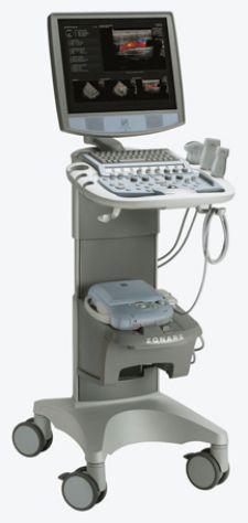
An impressive newcomer to the market, Zonare Medical Systems was recently voted as the 2007 “Best in KLAS” for hand-carried ultrasound (HCU) vendor with the z.one ultrasound system. “HCU is the fastest growing segment within the overall ultrasound market and one of the fastest in all diagnostic imaging,” reported a KLAS marketing research report. At the 2008 Annual Vascular Imaging meeting, Zonare highlighted enhancements to the z.one system that include a wider field of view on all linear transducers to allow clinicians to view more anatomy and pathology information on a wide range of patient types. The platform has also added the DICOM Structured Reporting for Vascular Exams feature, designed to automatically transfer calculation results to a PACS reporting system.
In May 2008, Siemens introduced the ACUSON P50, a laptop-based system with portability throughout the hospital and the capability to scan, process images, generate reports, view eMails and access the Internet. Siemens has also disclosed information about a new hand-held device called the Acuson P10, a Blackberry-size device weighing 1.6 pounds with a black and white monitor, flip-up liquid crystal display and two-dimensional mode imaging. This device is intended for a quick “look and see” and it is the target for early clinical intervention points such as emergency departments, cardiology, obstetrics, intensive care units, ambulances and medical helicopters. It is not intended to replace traditional ultrasound technology, but rather be a complementary tool in the progression of care and diagnosis.
Texas Instruments has also announced a family of fully integrated analogue front ends for portable to high-end ultrasound equipment. The AFE5805 will specifically address the portable ultrasound market by offering small size, decreased power consumption and low noise features for manufacturers of ultrasound systems. Production is scheduled for June 2008.
Another portable ultrasound system is the MicroMaxx ultrasound, which offers high-quality images with speed and reliability while efficiently operating on either battery or AC power. The system boots up quickly, from “off” to scanning in under 15 seconds. The small footprint of the 7.7-pound device plus its wireless throughput offers the flexibility only hand-carried systems can offer.
Reimbursement Worries
Improvements in care and technological advances always come at a financial cost. In the U.S., the Deficit Reduction Act (DRA) of 2005 has had a negative impact on freestanding imaging centers and independent diagnostic testing facilities. Reimbursement for portable ultrasound imaging may present a challenge. According to Eyal Herzog, M.D., a cardiologist at St. Luke’s-Roosevelt Hospital Center in New York City, “There is reimbursement for regular ultrasound, but there will be no additional reimbursement if you use small echo machines. On the billing code you can use 2D imaging, but you can’t bill for Doppler. I think they are working on this [reimbursement]. You can bill if there is color flow, but you cannot bill for Doppler.”
Even though some technological and reimbursement hurdles need to be addressed, hand-held portable ultrasound devices are promising valuable aids in clinical decision-making and they are less expensive than high-end machines. While portable units do not replace some of the applications of the high-end equipment, hand-held units are useful in a variety of settings.
Widening Range of Applications
Traditional areas of application have been prenatal care, radiology and cardiology, but additional settings and applications are expanding.
OB-GYN applications have ranged from in-hospital use, to physician office settings, rural hospitals and developing countries. In a recent clinical case, a visiting physician performed an ultrasound on a pregnant woman and determined that she was 32 weeks pregnant, not 40 weeks as previously reported. The timely and accurate diagnosis made a big
difference in this case, avoiding premature induction of labor.
In pulmonary medicine, ultrasound has been used for detection and quantification of pleural effusions, ultrasound-guided thoracentesis, lung tumor visualization and biopsy procedures and bedside assessment of lung pathology associated with severe pulmonary illness. Diacon and colleagues state*, “bedside examination is one of the advantages of ultrasound…at no time of day should there be a hesitation to take the machine to the bedside where it is needed.”
In the intensive care unit, bedside examinations are common and obviate the need to transport critically ill patients with attendant risks. The focused assessment for sonographic examination of the trauma patient (FAST) can assess fluid in the subxiphoid region for the pericardial sac, the right upper quadrant in Morison’s pouch, the left upper quadrant in the splenorenal recess and the pelvis in the pouch of Douglas or rectovesical space. The thorax can also be visualized for the presence of pleural effusions or hemothoraces and ultrasound can be further employed for guided thoracentesis. Additional examples of ICU applications include abdominal assessments, ultrasound-directed needle aspirations, gallbladder visualization, diagnosis of biliary obstruction, vascular access and DVT surveillance.
Applications of portable ultrasound devices in emergency room settings are most valuable for fast, accurate screening and triage decisions. Examples include FAST (focused abdominal sonography for trauma), immediate insight into critical patients, visualizing cardiac activity, obtaining information before ordering additional tests and improving bedside monitoring.
In cardiology, the clinical literature cites use of hand-carried ultrasound units as an extension of physical exam for estimations of EF, LV hypertrophy, regional wall motion abnormalities and pericardial effusion. Portable ultrasound units have influenced changes in medical therapy, diagnostic workup and decisions about cardiac catheterization or pericardiocentesis. While the hand-held devices may not have the same full capabilities of the traditional high-end machines, they offer an assist in bedside management.
“Portable ultrasound is used in the acute emergency and for suspected heart failure,” indicated Roxy Senior, M.D., DM, FRCP, FESC, FACC, professor, consultant cardiologist, director of cardiac research, Northwick Park Hospital (Middlesex Harrow, UK). “The portable system used at present has all of the features required to exclude significant heart disease.”
In terms of future desirable modifications, Dr. Senior noted, “There are now palm-held machines that will need harmonic imaging, basic Doppler and color flow and some form of storage.”
In a recent case, Dr. Senior used portable ultrasound for a patient with a life-threatening condition. “Recently, a patient presented to acute and emergency with severe shortness of breath,” said Dr. Senior. “Portable echo clearly showed a large accumulation of pericardial fluid. It is a life-threatening condition. In the absence of portable echo, we would have had taken a chest X-ray, which, with a pick-up rate of 80 percent, would alert the doctor to have an early echo. But even then, the patient would need to be transported to the echo department — all prolonging the diagnosis and potentially fatal.” In another case where a patient presented with shortness of breath, Dr. Senior used an echo to promptly rule out cardiac pathology and allowed physicians to quickly pursue other causes of shortness of breath, which in this case was lung disease. “The usefulness of a portable system is prompt diagnosis at the bedside, early management and early discharge,” said Dr. Senior. “Furthermore, it reduces unnecessary normal echoes to be performed in the main echo department; thereby reducing the waiting time for useful echoes.”
Other cardiac applications for the hand-held P10 portable ultrasound device include evaluating patients who do not fall into traditional risk categories, screening and identifying difficult-to-diagnose conditions such as left ventricular systolic dysfunction and left atrial enlargement, and identifying life-threatening cardiac issues in triage environments. Dr. Herzog participated in beta testing of the P10 device and commented, “We tested the design of the device by participating in a feasibility trial to see if the quality of the imaging was similar to the regular echo machine, and we were very surprised because the imaging was outstanding. We compared the data side by side to other imaging blindly, and we showed similarity in the quality of the imaging. The major advantages are that the P10 is small, portable and you can carry it in your pocket. When you do have a cardiac emergency situation, which happens not only daily or hourly in the CCU but with things happening every few minutes, you can immediately assist the patient. Since this is a clinical trial not clinical use, every patient we have here is compared to other imaging. But, we’ve had patients with cardiac tamponade, and patients with wall motion abnormalities due to acute heart attacks which we can diagnose.”
He added, “Until this type of portable device has color flow, it will not replace traditional echo machines because you cannot access valvular leakage. There are things you can identify without looking for valvular abnormalities, which include cardiac effusion or wall motion abnormality. Due to its size, feasibility and availability, it is to your advantage to use this device.” He added, “I do believe that in a few years we will be able to carry this in our pockets. In one of our drafts we call it echoscope, because you can look at the heart, not just listen to it.”
As the technology miniaturizes and ultrasound becomes more portable, the applications and utility of ultrasound are likely to expand, ranging from basic diagnostics, similar to that of a stethoscope, to assessing serious trauma such as heart attack, stroke or other life-threatening conditions.
*Reference: Diacon AH, Theron J, and Bolliger CT. Transthoracic ultrasound for the pulmonologist. Curr Opinion in Pul Med. 2005: 11; 307-312.

 May 07, 2026

 Clearance")


