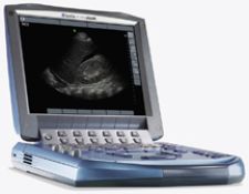
Sonosite's compact MicroMaxx 942 expands radiology services to areas it previously could not go.
They say that good things come in small packages, and if it has high-quality visualization tools, it’s even better. Medical imaging devices are miniaturizing and, as a result, opening up tremendous potential for taking advanced technology to the bedside.
While imaging modalities are becoming increasingly lightweight and portable, their features and performance are anything but small. Manufacturers are able to miniaturize existing diagnostic technology in ultrasound, CT systems and endoscopes, while still delivering robust computer power and memory capacity for advanced 3-D and 4-D imaging at the patient’s bedside.
Mini-Ultrasound Systems
One technology that is increasingly being miniaturized is ultrasound. Compact hardware allows for maneuverability in small clinical settings, portability to multiple sites and to the patient’s bedside. The smaller, portable devices enable clinicians to make pivotal patient care decisions using high-diagnostic quality imaging anywhere in the hospital or clinic, leading to improved patient outcome.
“The difficulty for a physician that goes to five different hospitals is that they may have five different ultrasound machines in the labor and delivery or prenatal area, all of which are mostly awful. The machines trickle down to the OB and they are the ones they don’t want anymore in radiology. And you can’t go down to radiology to use the machine at eight or ten o’clock at night that is four floors away,” explained Greggory DeVor, M.D., who delivers primary obstetric care as a physician consultant for 400 obstetricians and five hospitals in the greater Los Angeles area. The question for physicians is how to make critical decisions at multiple sites with adequate equipment.
Advantages of Compact Equipment
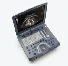
Instead of reinventing the wheel, many manufacturers simply miniaturized their existing ultrasound technology. GE Healthcare, for example, condensed its LOGIQ, Vivid and Voluson console systems into portable laptop versions like the Voluson e, equipped with labor and delivery, gynecological applications and assisted reproductive medicine. Both the Voluson e and Voluson i are designed to address one of the biggest challenges OB/GYN physicians face in caring for their high-risk patients – access to complete, real-time information that allows them to make clinical decisions at the patient’s bedside.
“I take the Voluson i with me and I have the same high technology that I have in my office. I can take it to the bedside because it is as big as a laptop. I can perform high-level studies and get maximal information,” said Dr. DeVor. “I have taken it to the home of patients who are home bound. It opens up tremendous potential for taking high technology to the bedside or wherever the patient may be.”
But does smaller hardware compromise technology? DeVor acknowledged, “There are some features that are not on the Voluson i that are on my other system, but those are very subtle features that are not required for labor and delivery. If I compare it to the image quality of the [larger ultrasound] that we use in the office, this is a nine and a half.”
Despite the laptop’s reduced screen size Dr. DeVor insists “that is why the laptop-sized screen is very sufficient for 95 percent of what we do.”
Portable Cost Savings
Portable units also benefit physicians who travel to several offices. Instead of having to buy multiple ultrasound machines, left idle while the office is not in use, the portability of ultrasound allows a physician to invest in just one system.
“They end up buying one machine and you take it with you and you have portability to go to the other offices. You can do that within your own office, if you put ultrasound laptop on a small cart and bring it into different exam rooms. It makes everything a lot easier,” noted Dr. DeVor.
Even within the same hospital, physicians need portable equipment. One solution is the hand-carried ultrasound by SonoSite, the TITAN. According to John S. Pellerito, M.D., chief of Division of Ultrasound, CT and MRI at North Shore University Hospital in Manhasset, NY, compact ultrasound has expanded the practice of radiology at his facility by extending services to areas that ultrasound previously could not go.
“I now have the ability to utilize ultrasound throughout the hospital. I can take a hand-carried unit wherever I need to examine a patient, and get a reading that directly impacts the treatment the patient is given,” said Dr. Pellerito. “In this way, the radiologist’s work is performed at the time of greatest need, and in the most convenient location.”
Compact Cardiac Imaging
In the emergency department (ED) where many patients present with chest pains, there is little to no time to carry that patient to another floor to diagnose a potential heart attack. To address the urgency of diagnosing cardiac conditions, GE Healthcare’s Vivid i is a miniaturized cardiovascular ultrasound system that provides high-performance, full-featured imaging in a lightweight design. Its wireless functionality enables physicians to consult at the patient’s bedside so that they can be more informed and involved in their healthcare decisions.
Siemens Medical Solutions’ answer to lightweight, fully featured, cardiac ultrasound systems is the X500 system. Miniaturized from Siemens’ top-of-the-line ultrasound platforms, this system is designed for a range of radiology and cardiology applications and offers optional transesophageal and stress echocardiography capabilities, and is available with fourSight 3D/4D imaging and the syngo Arterial Health Package.
USB Port Provides High-End
Image Access
Possibly the most portable device in ultrasound is the recently FDA cleared InnovaSound USB Ultrasound System by Direct Medical Systems, LLC. It is a Plug and Play ultrasound probe system equipped with standard ultrasound probe capabilities. The computer requirements for the USB ultrasound system are USB 2.0 and an operating system of Windows XP or Vista with a minimum of 1 Megahertz processor, 512 memory. This technology results in affordable ultrasound being available in nontraditional locations.
While the system has applications in ophthalmology, obstetrics and gynecology, otolaryngology (ENT), proctology specific to prostate care and diagnosis, emergency medicine, and vascular surgery, it is already destined for use in physical rehabilitation, where it is a component to a bio-feedback system that visualizes muscles contracting in real time.
Mini-CT Scanner Gets Around
Head trauma is another common emergency condition that requires immediate triage when a patient presents. However, wheeling a patient to the CT scanner can cause delays that result in permanent damage. To address the immediacy of having a CT-scanner on hand, Neurologica Corp. developed the CereTom CT, a portable, wireless system that generates up to eight slices per revolution, using a Modular Multi-Row Detector (MMD), and completes a full neurological scan in reportedly 30 seconds. “The device can be brought to the patient’s bedside in the ICU or other locations, reducing the time and dangers associated with moving a critical patient to the radiology suite,” said Neurologica Corp. President and CEO Eric Bailey.
Although the CereTom delivers the same resolution as large CT systems, the scanning speed is slower. But Bailey says the scanning speed for heads for the CereTom is not substantially different from many of the standard head protocols utilized in the larger CT machines. For a non-contrast head scan, an international supplier utilizes a standard protocol of 170 milliamperes at 4,000 milliseconds per rotation as compared to the CereTom’s standard head protocol of 8 milliamperes at 4,000 to 6,000 milliseconds per rotation.
Mini-Endoscopes Get the Big Picture
Another area where mini-imaging devices are capturing the big picture is in gastroenterology, in particular with capsule endoscopy.
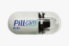
The newly FDA-cleared PillCam SB 2 video capsule by Given Imaging is a tiny endoscopic camera that the patients swallow. The device takes snapshots inside the gastrointestinal tract. The advantage of the video endoscope is that it visualizes areas that may be overlooked by conventional endoscopy, in particular in the small intestine.
One image-enabled endoscope currently being tested “swims” through the GI tract. Developed by Nobuhiko Hata, the technical director of image-guided therapy at Brigham And Women’s Hospital and a professor of Radiology at Harvard Medical School, this image-snapping robot has free-style maneuverability as it swims through the GI tract propelled by fins that are magnetically guided by an MRI system. This is unique because current capsule endoscopes are passive or simply row in a straight path through the tract.
As imaging devices miniaturize, the clinical view broadens, opening up tremendous potential for high-quality imaging for any part of the body, from any location.

 May 07, 2026

 Clearance")


