Various definitions for artificial intelligence (AI) have emerged as the technology has evolved. Approximately 20 years ago, the late John McCarthy, an American computer scientist credited with being one of the founders of the discipline, defined AI as “the science and engineering of making intelligent machines, especially intelligent computer programs. It is related to the similar task of using computers to understand human intelligence, but AI does not have to confine itself to methods that are biologically observable."
Today, in its simplest terms, AI is still intended to teach machines how to think and learn independently while performing tasks that typically require human intelligence, such as understanding language, recognizing patterns, solving problems and making decisions. The world is observing how AI can transform a variety of industries, including those that rely on image processing to provide patient care services, such as radiology, the science of diagnosing and treatment of diseases by employing X-rays and other high-energy radiation. Just as with previous advancements through image archiving and magnetic resonance imaging, AI is reshaping this healthcare practice through innovative tools and technologies that augment the capabilities of healthcare providers while streamlining workflows and enhancing diagnostic accuracy.
AI for Radiology: An Evolving Science
The first reports of AI’s use in the radiology field date back to 1992,1 when it was utilized to detect microcalcifications in mammography or computer-aided detection (CAD). Although the reputation surrounding CAD in this industry has not been particularly strong over the past four decades, with studies determining that its presence has reduced radiologists’ accuracy and offers no benefit to patients, this trend appears to be shifting. According to a 2019 report by the Radiological Society of North America (RSNA), when used in collaboration with deep learning, a subset of machine learning (ML) in which the computer network teaches itself to understand concepts without human involvement by performing calculations on a large dataset, AI is essentially “re-inventing” the CAD process by improving efficiency and reducing the incidence of “false alarms.” Deep learning is the most popular category of AI in radiology and uses sophisticated neural networks to detect patterns within input data to produce outputs. For example, according to a recent study,2 “deep learning can learn the relationships between pixels in a chest X-ray to detect findings associated with the presence or absence of pathologies, such as cardiomegaly, emphysema and atelectasis, at a level similar to practicing radiologists.”
Introduction of Radiomics
As AI has evolved, the concept of radiomics, a rapidly growing field of research concerned with the extraction of quantitative metrics within medical images, has emerged. Radiomic features have quickly become a reliable way to capture tissue and lesion characteristics, such as heterogeneity, that can be used for clinical problem-solving when used alone or in concert with demographic, histologic, genomic and proteomic data. Radiologists can also interpret these digital images in a qualitative way that can then be transformed into quantitative data. Rather than automation, radiomics builds upon already available data for a given imaging study through quantification of the spatial distribution of signal intensities and the pixels/voxels of the 2-D or 3-D study, thereby functioning as an additional tool to aid clinicians in various diagnostic and prognostic clinical decision-making.
Radiomics also helps to highlight subtle differences in image intensity, geometry and texture, thereby overcoming the limitations of the human eye. Predictive data analytics and pattern recognition aid in the early detection of various pathoses, such as the distinction between benign and malignant lesions, thus influencing treatment planning and the performance of treatment regimens.
The rise of AI and ML algorithms has led to the development of statistical natural language processing (NLP), a branch of AI that gives computers the ability to understand and interpret verbal human language and written text. According to RSNA, NLP has developed into an invaluable tool with significant potential in radiology, particularly related to the analysis of reports and research through open-source libraries. Radiologists who understand the potential and limitations of NLP will be better positioned to evaluate how the science can improve clinical workflow and facilitate large amounts of research.
Furthermore, more AI applications are now utilizing large language modeling (LLM), a deep learning algorithm that enables computers to recognize, translate, predict or generate text and other content. LLM is changing the landscape of NLP by offering possibilities to transform treatment development more effectively by being applied to report creation, speech recognition, summarization, and text classification. Suitable examples of LLM usage include the safe extraction of data from existing radiology reports without compromising patient privacy and improving the overall efficiency of report writing LLM models can also be applied to generate prompts based on patient symptomology and response to therapy and probability analysis from large patient datasets. In contrast to ML, the comprehension and generation of free-form text by LLMs are employed in this application. Medical diseases and conditions for which large volumes of data exist through longitudinal and cross-sectional studies further benefit from LLM-based digital assistants that can provide daily patient recommendations and guidance, and identify situations that might require physician intervention.
AI’s Impact on Patient Care
AI can be applied to almost every part of the patient journey, including scheduling appointments, selection of examinations, insurance transactions, imaging appropriateness and reporting workflow (see Figure 1). Currently, the U.S. Food and Drug Administration (FDA) has authorized more than 500 AI-based medical devices. Various AI tools have also been integrated into clinical decision support software, image viewing software and medical dental record software systems to improve care. A 2021 study3 released a medically aware GPT-3 data labeler, known as the GPT-3-ENS, which combines medical information with GPT-3 (open AI) for precise medical dialogue summarization.
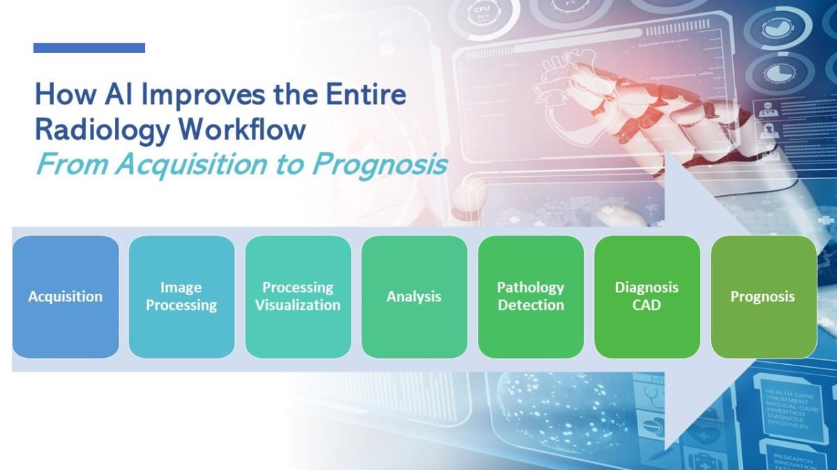
Figure 1: How AI Improves Radiology Workflow
Due to the aging population and a greater reliance on imaging in the United States, the use of computed tomography, magnetic resonance imaging and ultrasound imaging has dramatically increased over the past two decades. AI-based bone age estimation systems, such as the BoneXpert, launched in 2009, have been reported to reduce reading time by up to 40 percent.4 Another example of AI’s benefits is a reduction in reading time by more than 20 percent5 in the detection of pulmonary metastases as well as the widespread usage of AI software for the detection of tuberculosis on chest radiographs.
Upcoming Challenges
Although the science of AI has displayed encouraging promise and the technology is developing a ubiquitous presence, the field is still in its nascent stage and experiencing multiple challenges. These include the desirability for technique standardization, lack of sufficiently large datasets, and extensive research studies that affect the reproducibility and generalizability of findings, accurately accounting for various patient variables that impact radiomic feature values, and the relatively high risk of false-positive results on imaging studies. Furthermore, it is important to address the ongoing privacy and data security concerns inherent with advanced technology being implemented into healthcare as widespread implementation and adoption occurs.
Recent guidelines aim to facilitate quality control and standardization within the field. The Radiomics Quality Score (RQS), a number that assesses the characteristics and quality of radiomics studies and the reports they generate, is calculated by evaluating preset criteria.6 The transparent reporting of a multivariable prediction model for Individual Prognosis or Diagnosis (TRIPOD) checklist also operates through a preset model meant to predict the effectiveness of individual prognostic or diagnostic conclusions.7
The school of AI is not limited to specific fields of expertise. Wherever a particular problem exists about the processing of an image, consumption of an image, or any remote processing of images — AI can help. At the same time, however, there is a certain underutilization of AI occurring based on the breadth of its capabilities. Industry professionals can be enlightened on how to better use their tools moving forward. Various radiology education institutes offer AI-oriented fellowships and training programs to help prepare radiology technicians and clinicians to incorporate AI into their daily workflow. As the debate about AI being a bane or a boon continues, various seminars are being held to raise awareness of how AI can be a valuable tool for assisting instead of replacing the clinician’s role in patient care.

Akshay Chaand is an IT product manager, software developer, electronics engineer and subject matter expert with a strong background in healthcare, construction tech, integrations and Dev/QA software development. With more than 14 years of experience in technical product management, agile leadership and cloud engineering, he is also skilled in market research, product roadmap development and data analysis. He holds a master’s degree in VLSI and microelectronics engineering from Kurukshetra University.

Meeru Kumar, BDS, MSc (Oral Medicine), PG Cert (Oral and Maxillofacial Radiology) is a practicing dentist with specialty training in oral medicine and oral and maxillofacial radiology from Texas A&M School of Dentistry. She has extensive experience in clinical dentistry, radiographic imaging techniques, diagnostic sciences, and clinical research. Kumar is passionate about advancing medical technology and patient-focused healthcare.
References:
- https://ascpt.onlinelibrary.wiley.com/doi/pdf/10.1111/cts.12704, accessed Dec. 12, 2023
- https://mededu.jmir.org/2023/1/e43415, accessed Dec. 12, 2023
- https://aclanthology.org/2021.nlpmc-1.9/, accessed Dec. 12, 2023
- https://pubmed.ncbi.nlm.nih.gov/28898126/, accessed Dec. 12, 2023
- https://pubmed.ncbi.nlm.nih.gov/32037256/, accessed Dec. 12, 2023
- https://pubmed.ncbi.nlm.nih.gov/36282312/, accessed Dec. 12, 2023
- https://pubmed.ncbi.nlm.nih.gov/31350588/, accessed Dec. 12, 2023

 July 20, 2026


