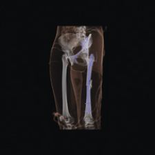
This image of the pelvis and femur was obtained for post-op follow-up and shows the details of not only the bones but also the metal plates, rods and screws. The image was acquired on Siemens Medical Solutions' SOMATOM Sensation 64 with z-Sharp technology.
Today patients present to the emergency department (ED) with a slew of complaints and injuries ranging from a chronic cough to a sudden life-threatening head trauma. As such, the ED is no longer reserved for those patients with dire life or death circumstances, as the standards of emergency care apply to all patients and handle a larger caseload.
With this changing dynamic, physicians rely heavily on imaging modalities to quickly triage patients and uphold emergency standards of care in providing an accurate, complete and immediate diagnoses. In many respects, the ED is becoming more of an emergency radiology department or ERD.
Radiology Takes Over the ED
A leader in the ED to ERD trend is Brigham And Women’s Hospital, an affiliate of Harvard Medical School. Stephen Ledbetter, M.D., chief of the Emergency Radiology Division at Brigham and Women’s, explains that “a dramatically increased use of the emergency department by patients and a growing volume” has led to the creation of their emergency radiology division. “That trend is going to continue because of demographics — which includes an aging, more diverse and uninsured population.” Because of this increased use, he explained, “Emergency medicine has become a full-blown specialty just like neurology or plastic surgery, and like other specialties it now needs services that are coordinated and aligned with its clinical service. Emergency medicine needs a dedicated emergency radiology section to ensure the smooth delivery of care. Emergency radiology is a different vehicle for the delivery of emergency medicine services. It is no longer a delivery vehicle that tries to obtain services from five, six, seven or eight subspecialties of radiology. It needs those subspecialty services coordinated under one umbrella.”
Specifically, an ED develops into an ERD once it is delivering emergency radiology coverage 24/7 and has around the clock access to imaging tools to support it. “Historically, emergency radiology was on-call radiology and that’s why, in the past, there was no emergency radiology department. The old model included extended hours, multiple consecutive hours and working alone with limited resources. There was not a dedicated section to provide and foster oversight, advocacy and planning. Emergency radiology takes that historic model and flips it on its head; it has moved from an on-call model to a service model,” according to Dr. Ledbetter.
The ED provides around the clock radiology coverage by having constant access to the latest technological advancements in imaging. Although one may have equal access to computed and digital radiography (CR/DR), three-dimensional computed tomography (CT) and magnetic resonance (MR) imaging , several factors come into play when determining which modality is best suited for a given patient with a given medical condition. While CT and MR provide the best imaging results, CR and DR are the most frequently used for patients presenting in the ED because these systems are less expensive, expose patients to the least amount of radiation and are fast.
Commenting on the convenience of CR and DR, Steve Nokes, M.D., chief of Radiology at Baptist Medical Center in Little Rock, AR, stated, “In fact, in our hospital, radiologists read and put a voice clip on the CR/DR images so that anyone can hear that from any workstation. That’s a world of difference from just two years ago for us. X-ray would come over, we would dictate, then phone in to the dictation system. Now you just look at the image of the chest.”
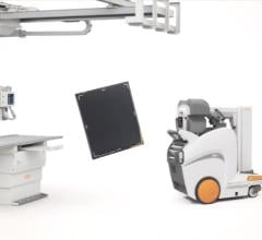
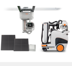
Blaine Morris, MBA, vice president of Clinical Services at Erlanger Medical Center also expounds upon the reasons for the primary use of CR/DR imaging tools in the ED. His hospital sees roughly 60,000 level one trauma patients per year. With the heavy volume of patients, his ED relies heavily on portable CR and DR machines, including the Kodak Directview DR 9000, the CR 975 and the CR 950 by Carestream Health Inc. Morris explains that a typical work up on a given patient usually includes one of three things – a cervical spine X-ray, chest X-ray or a pelvic X-ray. “Those are all on CR/DR,” noted Morris. “They are doing those concurrently with blood work. They can be intubating while they’re doing the CR/DR. Now, it’s not just one or the other. Instead, you can have care customized as they [patients] come in the door. It’s really incredible all the things that can be done at the same time.” One of Carestream’s latest products includes the CR-ITX 560 system, which combines a powerful CR reader and a transportable X-ray unit in a single, self-contained package. It is designed for convenience, enabling immediate capture and access to patient images where time is critical for accurate diagnoses.
Accuracy, Specificity Drive CT and MR in the ED
With the heavy reliance on CR/DR for many indications presented in the ED and with its convenience, quick assessment and low cost, what then prompts physicians to order CT or MR? According to C.E. Ballard, M.D., emergency department staff physician at Baptist Medical Center in Little Rock, AR, “The chief complaint for a plain CR/DR image is an indication of an acute, chronic or exacerbation of a chronic illness, a chest X-ray or a long bone X-ray. If we get that X-ray and it’s unclear, then frequently the radiologist may indicate a CT or MR.” Similarly, William R. Reinus, M.D., MBA, FACR, vice chairman of the Department of Radiology at Temple University Hospital concurred, “If we can’t figure out what’s wrong, it’s better to spend money upfront than have something disastrous occur. With the legal repercussions that exist, most people are inclined to be fairly liberal in the number of studies we order.”
Dr. Ledbetter also realizes the potential of CT, calling it a “close second” to CR/DR use. “The real driver behind the increased use of CT is the improvement in its technology,” he said. “In addition, the need for increased sensitivity and increased specificity during any visit is a major trend in emergency care. That’s at odds with decreasing utilization. The reason for the growing use is two-fold: Improvements in technology are making it a better exam for evaluating patients, and at the same time there is the growing pressure for increased sensitivity for the ability to detect disease and specification – the ability to say that this is abnormal, but this is the specific diagnosis.”
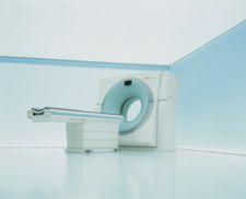
Because of the advancements in technology, specifically in CT, Dr. Reinus uses Siemen’s SOMATOM Sensation, a 40- and 64- slice CT using the latest z-Sharp technology, which provides the highest routine isotropic CT resolution of 0.33 millimeter voxel size and complete elimination of spiral artifacts. Siemen’s SOMATOM Sensation is able to scan a thorax in four seconds and freeze the heart’s motion using the industry’s fastest gantry rotation time of 0.33 seconds. According to Dr. Reinus, “The goal that we need to work toward is to get the maximum amount of imaging with the least radiation exposure. To that end, it’s certainly been the progression of CT to do more, faster, but at some point people are going to have to think about doing survey-projectional images and cross-sectional images. The patient will need be in the scanner and need a topogram in different planes in which the patient only goes through the scanner once with contrast and once without.” CT has become quicker and more accurate, however, each new advancement and improved efficiency prompts one to dream even further into the future of imaging.
CCTA Becomes Critical in the ED
Another imaging tool on the cusp of more widespread use is coronary CT angiography (CCTA). It shows promise in its ability to rapidly and accurately assess chest pain by providing an inclusive set of images for evaluating the three main causes of chest pain: a pulmonary embolism, aortic dissection and coronary artery stenosis. The CCTA, using the latest 64-slice multidetector computed tomography (MDCT) systems, enables visualizing the entire chest of all three areas of suspicion. This type of assessment can be performed in about 12 seconds; however, the 256-slice system that is currently being tested in Japan can scan the heart in 1.5 seconds.
Although ultrasound has been widely used for many years, researchers continue to explore new applications for it. According to researchers at Yale University, ultrasound is able to screen for abdominal aortic aneurysms (AAA). Thirteen of the 179 patients in the Yale study were found to have AAAs, the sizes of which averaged 4 cm (with a median of 3.7 cm). In those patients, the average time to complete the study was 141 seconds (with a median of 138 seconds). So far, 10 of those patients have received formal imaging in follow-up, and all had AAAs with an average discrepancy of 0.6 (median 0).
MR Sets Gold Standard, Yet not Fast Enough
With all of the focus on CR/DR, CT and ultrasound, where does MR come into play? According to each physician interviewed, even though MR has exceptional image quality, it is the least utilized in emergency situations. The main reason for this lag in utilization of MR is that it takes too long. Dr. Nokes indicates that MR is used to diagnose strokes in the ED. According to a recent study conducted by the physicians at the National Institute of Neurological Disorders and Stroke (NINDS), MR was found to outperform CT in the emergency diagnosis of suspected acute ischaemic stroke. MR was found to be about five times as sensitive as immediate noncontrast CT and twice as accurate as CT. Noncontrast CT and MR were found to be equally effective.
Despite the recent findings, Dr. Nokes explains that a patient suffering a stroke has only a three-hour window from the time of the stroke to receiving therapy. Most of that window is lost by the time the patient realizes they have had a stroke and reach a hospital. According to Dr. Nokes, MR takes too long in these situations even though it may outperform CT. For patient management, in a hospital with a stroke center, MR has not taken over the role of aggressive anti-coagulation of that patient either by intravenous or interarterial thrombosis in diagnosing a stroke. “CT does a really good job of seeing blood in the brain. We do a MR soon after a CT to show that there is a stroke,” said Dr. Nokes.
According to Dr. Ledbetter, “MR is a great tool if it weren’t for the fact that the exam times are longer than we would like in an emergency care setting and access is an issue during the after hours when patients are seen. CT is immediately available and staffed 24 hours a day. Every ER has at least one, if not a fleet of CT machines. As access to MR improves and more radiology practices organize an emergency radiology section, they will be able to access those modalities because they will be able to work on exam times and advocate for staffing resources that they need. I expect that in five to 10 years there will be a significantly greater use of MR as well.”
What is clear about the future is that advanced imaging using CT and MR will play an increasingly important role in accurately triaging trauma patients.
 January 08, 2026