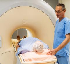
PET/CT allows clinicians to precisely target tumors.
Visualizing motion is changing the way oncologists and medical physicists treat cancer patients. Thanks to multislice CT and advanced post processing, clinicians can see tumor movement as a result of respiration clearly and precisely with new 4-D imaging techniques.
Changing treatment one patient at a time
As one of the nation’s largest integrated community networks of cancer physicians and specialists, UPMC Cancer Centers offers the latest technology for cancer care. This premier National Cancer Institute-designated comprehensive cancer center has a basic premise – no patient should have to travel more than 20 minutes for cancer treatment. More than 40 locations throughout western Pennsylvania, covering a geographic area of more than 100 miles around metropolitan Pittsburgh, fulfill this vision. The centers are linked by a telecommunications network that enables patients in rural areas outside Pittsburgh to access state-of-the-art radiotherapy and imaging services typically only available at large academic medical centers.
Dwight Heron, M.D., associate professor and vice chairman of Clinical Services at University of Pittsburgh Medical Center (UPMC) Cancer Centers has used 4-D CT imaging for radiation therapy planning for the past three years. “The benefits of 4-D CT are significant, especially with tumors in the lung or upper abdomen such as the esophagus, pancreas or liver,” Dr. Heron said. “With respiration, structures around the tumor may move. With 4-D CT, we can actually see the tumor or organ move.”
Thanks to this advanced imaging technology, Dr. Heron and his associates have a true representation of a patient’s respiratory cycle and the impact on tumor movement. From this data, the clinician can determine the degree of movement and create a treatment plan during specific phases. “We can minimize excursion during a particular phase to decrease margins around the tumor and as a result lessen toxicity to healthy tissue and nearby organs,” he added.
As an example, treatment plans for lung tumors typically have larger margins due to respiratory motion. With vital organs surrounding the area, clinicians also typically limit the radiation dose for fear of treatment toxicity. Dr. Heron ponders if this “may be the reason we haven’t been able to control lung tumors to the degree we would like.” He does believe 4-D CT as well as newer treatment techniques hold much promise for combating this specific disease.
For treatment, UPMC uses Intensity-Modulated Radiation Therapy (IMRT) along with 4-D gating. “We can also escalate the dose without injuring the surrounding tissue,” Dr. Heron explained, “and increase the local control rate, while actually reducing toxicity.” Without 4-D gating, Dr. Heron would have to limit the dose to avoid toxicity. “4-D CT enables us to make better treatment decisions.”
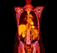
While over 450 patients have been scanned with GE Healthcare’s Lightspeed Plus wide bore CT simulator and 4-D CT application, UPMC hasn’t stopped there. Applications like AdvantageSIM MD on the oncology workstation have let clinicians fuse multimodality images such as 4-D PET, since UPMC began capturing 4-D PET images in the fall of 2006.
“We’ve taken it one step further with 4-D PET and now have the PET image overlaid with 4-D data to see both anatomy and function,” Dr. Heron explained. “This provides a wealth of data that is absolutely amazing.”
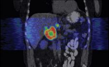
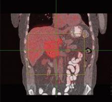
Dr. Heron cites a specific clinical example of 4-D CT/4-D PET imaging’s impact on patient treatment planning. In a patient with an upper abdominal mass, Dr. Heron could not conclusively tell exactly where the tumor was on a typical helical CT scan. He could see the tumor with PET. However, once he fused the images and created a 4-D PET/CT data set, Dr. Heron could identify nearby lymph nodes that weren’t obvious on the helical CT, 4-D CT or PET scans.
“This information dramatically changed our treatment plan for the patient. With the 4-D data, we learned the center of the tumor was hypoxic, and, therefore, we could use that knowledge to deliver more radiation to the center of the tumor,” indicated Dr. Heron.
By using a timed resolution, UPMC can fuse and link together 4-D PET and 4-D CT images to precisely view tumor movement. “It is no longer sufficient to capture only 4-D anatomic images.” Dr. Heron explained, “It’s a natural evolution to also have images depicting 4-D function.”
Thanks to the advanced technology at UPMC, 4-D data shows that in over 95 percent of patients, as the tumor shrinks during treatment, its trajectory is almost identical to that which was mapped prior to the treatment. “With our data, we are confident that even as a tumor shrinks, it still moves in the same way. This knowledge now opens the door to greater possibilities in improving cure rates and decreasing side effects of cancer treatments with radiation for our patients.”
How to track a moving target
There is a wide range of tools available for clinicians to precisely track tumors. GE Healthcare introduced 4-D CT four years ago, and as a result of the company’s joint effort with Varian Medical Systems, GE added Varian’s RPM Respiratory Gating System to its CT simulators.
“Whether or not therapy delivery is gated, it is important for clinicians to know the exact location of a tumor as it moves through the respiratory cycle so they can treat appropriately,” said Kelly Piacsek, Ph.D., oncology marketing manager, GE Healthcare. “Once clinicians see how much tumors move with respiration, they realize they can’t treat without this imaging capability.”
Cine mode is a unique feature in 4-D visualization that allows retrospective selection of the very precise respiratory phases at any location, and GE offers the ability to acquire in cine mode. “This capability allows a nearly unlimited scan range and phase accuracy unattainable with helical scanning,” Dr. Piacsek added.
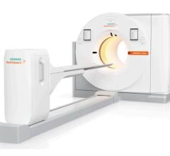
Accurately mapping out radiation therapy requires precise localization of a tumor, which is often a moving target. Kulin Hemani, vice president of Oncology Care Systems Group, Siemens Medical Solutions, agrees that the “knowledge of tumor characteristics, location and motion with respect to the breathing pattern provide clinicians with the information they need to create a very accurate treatment plan.” With SOMATOM Sensation Open CT the user benefits from Siemens’ unique STRATON tube, which has a large heat dissipation capacity. This allows clinicians to capture large volumes of data in one contiguous scan without the limitation of cooling delays. Breathing can cause a tumor to appear double in length or half its size. The ANZAI gating system may be used on both the Sensation Open and Siemens’ linear accelerators – optimizing treatment delivery by using the entire motion management process for the true size of the tumor.
Philips Medical Systems’ Tumor Localization (Tumor LOC) application helps localize target volumes for radiation therapy planning. It includes features for viewing Respiratory Correlated CT data sets and analyzing motion of target and surrounding anatomy. Until now, Tumor LOC has only been available to Philips Brilliance CT Big Bore customers on the console, but with software release 3.5, this application will be available on the Extended Brilliance Workspace (EBW). When offered together with Remote Reconstruction and Pulmonary Viewer, it turns the EBW into a high-performance 4-D oncology workstation, allowing visualization of respiratory motion from a beam’s eye view perspective.
The integrated multimodality workspace from Siemens includes, among other applications, 4D Inspace, virtual simulation and multimodality fusion software. 4D Inspace lets clinicians view tumor motion in 3-D. That data can then be transferred to any DICOM- compliant treatment planning system for accurate and effective radiation therapy. “By viewing the tumor motions in 3-D, clinicians can identify accurately tumor location and synchronize dose delivery with the patient’s breathing cycle,” added Hemani.
Similarly, GE’s Oncology Work-station and AdvantageSIM MD virtual simulation software features multi-phase, multimodality image management that also assists with RT planning. This system allows users to contour and plan on a movie loop of the tumor through the respiratory cycle. This allows the clinician to utilize all phases simultaneously and minimizes guesswork when planning treatment — which is half the battle.

 April 23, 2026