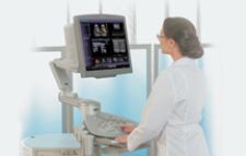
The new ACUSON Antares ultrasound system by Siemens Medical Solutions visualizes breast lesions using an elasticity imaging technology.
The same technology that was once used to measure distance under water today can visualize blood flow in the fetal heart – a testament to the evolution of ultrasound technology.
Ultrasound has consistently improved technologically in clinical diagnostics and therapeutics, and, recently, has broadened its applications in magnetic resonance-guided focused ultrasound surgery, ultrasound elasticity testing and drug-ultrasound stroke treatment.
Ultrasound’s Role Expands at Point of Care
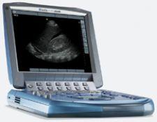
“With all of the advances, the development and evolution of hand-carried ultrasound has had the broadest impact in expanding the clinical applications of ultrasound by moving ultrasound out of the imaging lab and bringing it to point-of-care applications in acute and critical care in a variety of hospital and nonhospital settings,” stated Anne M. Bugge, vice president for Corporate Affairs of Sonosite Inc., which released its MicroMaxx hand-carried ultrasound system in April 2005 and has since installed more than 30,000 ultrasound systems in hospitals, clinics and physician offices worldwide. The MicroMaxx system weighs less than eight pounds and can withstand being dropped on a hard surface and continue to function, according to Bugge. “This combination of performance and durability plus its intuitive user interface makes it ideal for use in point-of-care applications where the ability to make quick assessments can be life saving and the need to provide visual guidance for invasive procedures can reduce risk,” said Bugge.
David Edrington, M.D., a board certified physician in Emergency Medicine at Baptist Hospital in Little Rock, AR, explains that he often uses Sonosite’s TITAN ultrasound system, which, similar to the MicroMaxx system, is compact, portable and easy to use. Describing the benefits of using the TITAN in acute care settings, he said, “I have central venous access immediately. Ultrasound gives me the option of having direct visualization. Radiologists don’t have time and are overburdened with interventional procedures. So instead of waiting for a radiologist, I can expedite patient care.” Edrington says that 90 percent of the time he uses the TITAN ultrasound system for vascular venous localization of internal jugular veins to place central lines, and the other 10 percent is used for localization in order to perform thoracentesis or paracentesis. For Edrington, Sonosite’s hand-held ultrasound systems make emergency medicine practice more efficient and cost-effective with obvious benefit to patients.
Another innovation in ultrasound that shows much promise is volume imaging. “Volume Imaging is the next revolution in ultrasound since it improves productivity and allows the viewer to manipulate the data and see planes that were not previously available. This improves diagnostic confidence,” said G. Parhar, director of Ultrasound Business Unit for Toshiba America Medical Systems. In November of 2006, Toshiba introduced its Aplio XG which features several enhancements, including volumetric imaging for radiology applications, such as 3-D multislice view, in which users can select slice thickness and the number of images displayed, and new 4-D technology that enables the user to obtain volume data sets for later off-line review. “We use the Aplio on all of our vascular imaging studies that we offer: carotid duplex; imaging of the arteria and venous circulation of the arms and legs; AV hemodialysis grafts; bypass grafts; as well as abdominal arteries such as the abdominal aorta, renal and mesenteric arteries,” said Megan Hodge, RN RVT, manager of the Vascular Diagnostic Laboratory, DeBakey Heart Center of The Methodist Hospital in the Texas Medical Center, Houston, TX.
Scanning the Fetal Heart in 3-D/4-D
Volume imaging has also had a significant impact on understanding the fetal cardiovascular system. Recent advances in the technology of 3-D/4-D ultrasound systems allow almost real-time 3-D/4-D fetal heart scans. Leeber Cohen, M.D., who specializes in obstetrics and gynecology ultrasonography at Northwestern Memorial Hospital in Chicago, IL, is planning research to investigate the new STIC (spatio tempural image correlation) technology to assess fetal hearts. Obtaining images of the fetal heart is challenging due to the heart’s rapid movement. However, STIC enables physicians to obtain multiple volumetric images of the heart and synchronizes them to the fetal heart rate. The resulting image is then displayed in real time with which the user can select various images for viewing. Cohen asserted that the obvious benefits of the new STIC technology include less ultrasound exposure, gated Doppler evaluation of the fetal heart and the ability to get second opinions by telemedicine. However, one downside of this new technology concerns image resolution.
“When a 2-D image is taken out of a volume, there is a loss of resolution, resulting in poorer image quality,” stated Cohen. He explained that there is no evidence supporting that 3-D imaging is better than 2-D imaging for routine screening; for now 3-D is best used when evaluating a known suspect anomaly. However, Cohen comments that this may soon change due to the development of a new probe.
Currently, mechanical probes are widely used. A new generation of matrix array probes based on 8,000-10,000 silicon-based microtransducers should be available in three to five years with much better image quality than either mechanical probes or the present generation of matrix array probes. Theoretically, the image quality with electronically steered matrix arrays should provide better image quality. With mechanical probes, multiple 2-D slices are obtained by a mechanical probe and stacked. The new generation of probes using matrix array technology will enable direct volume scanning by acquiring pyramidal volume sets and reducing motion artifacts.
Already available for clinical use is the Voluson E8 by GE Healthcare. The system includes sonography-based volume computer-aided diagnosis, known as SonoVCAD and an automated imaging tool. SonoVCAD is intended to simplify volume imaging of the fetal heart by automatically generating multidimensional images of outflow tracts after the standardized four-chamber view has been obtained.
Expansion in Breast, Stroke and Fibroid Treatments
To further boast ultrasound’s advancements in image quality, the new ACUSON Antares ultrasound system by Siemens Medical Solutions has made a breakthrough in visualizing breast lesions using an elasticity imaging technology. With elasticity testing, patients undergo a standard ultrasound to view the breast lesion initially seen on mammography. A second ultrasound is performed using elasticity imaging which further demonstrates the lesion’s rigidity and size. If the lesion is larger in the second screening, chances are that it is malignant. Preliminary results using Antares for breast lesion evaluation has demonstrated 100 percent specificity in correctly identifying cancerous lesions and has shown 99 percent specificity in identifying benign lesions. With 75 to 80 percent of breast biopsies believed to be unnecessary, this new technology may save women the time, money, pain and worry of undergoing invasive breast biopsies.
Another promising finding resulted from an investigation that evaluated the effectiveness of ultrasound in treating stroke patients. A pilot study demonstrated that ultrasound microcatheter thrombolysis, an application of low-energy ultrasound administered either transcutaneously or percutaneously through a catheter with an external ultrasound transducer at the site of the clot, in combination with an intra-arterial tissue plasminogen activator (tPA) reopened occluded arteries in stroke patients better than medication alone when administered within three hours of onset. Sound waves emitted by ultrasound assist in breaking up the fibrin, the foundation of a blood clot. This, in turn, creates more plasminogen activation receptor sites and expedites clot dissolution. In this trial, 69 percent of patients achieved complete reopening of the obstructed artery.
In another study, magnetic resonance-guided, focused ultrasound surgery, a noninvasive procedure, was evaluated for its ability to alleviate uterine fibroid systems. Before treatment, women received magnetic resonance imaging to identify the fibroids to be treated. Next, focused ultrasound was administered to supply heat to the fibroid which destroyed the tissue and immediately stopped the flow of blood within the tumor. The result was a significant decrease in uterine fibroid systems, such as excessive menstrual bleeding, frequent urination, pelvic pain and infertility for up to 36 months. This procedure could mean less need for hysterectomies, myomectomies and other invasive procedures, which currently are the norm for treating fibroids.
From improving treatment outcomes in patients to better diagnostic confidence for physicians, ultrasound continues to deliver cutting-edge technological breakthroughs for the advancement of medicine. Ultrasound’s trajectory throughout history, originating as a device for measuring sound to serving as one of medicine’s most relied upon imaging tools, suggests that it may shore up several more applications in the future as the technology continues to evolve.
Find the Ultrasound comparison chart online at www.ITNonline.net.

 May 07, 2026

 Clearance")


