News
Montage Healthcare Solutions, Inc., a provider of search-driven radiology analytics and data mining tools, announced that more than 27 healthcare organizations have added Montage Search and Analytics to their Nuance PowerScribe 360|Reporting workflow. By tightly integrating these solutions, radiologists can have more data in their hands to optimize decision-making during the dictation process. Montage Search and Analytics can be launched from within PowerScribe 360|Reporting, providing radiologists with clinical decision support through seamless access to historical reports.
 September 18, 2013
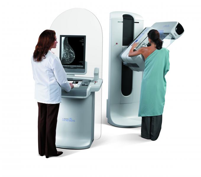
Feature
A new analysis has found that most deaths from breast cancer occur in younger women who do not receive regular mammograms. Published early online in Cancer, a peer-reviewed journal of the American Cancer Society, the study indicates that regular screening before age 50 should be encouraged.
September 18, 2013 News
Since 2007, John Muir Health’s Clinical Research Center has been participating in a large-scale research project alongside dozens of other hospitals around the world as part of the International Early Lung Cancer Action Program (I-ELCAP) and has screened 250 patients to date. The study quantifies the benefits of low dose CT scans for early lung cancer detection in high-risk patients – current and former heavy smokers ages 55 to 80.
September 17, 2013 Sponsored Content
Sponsored Content
Videos | Radiology Business
Radiology departments have many different needs and face a wide variety of challenges that can impact their departments ...
November 11, 2025 Technology
ClearCanvas introduces its new vendor neutral picture archive and communications system (PACS), Cleome, to its portfolio of commercial products. This Web-based breakthrough solution features fast diagnostic viewing of all medical images using zero-footprint HTML5, anytime, anywhere.
September 17, 2013 
Feature
A team of California-based breast imagers and breast cancer risk specialists have developed a website to help navigate the new challenges posed by breast density notification laws, according to a special report published online in the journal Radiology.
September 16, 2013 
Technology
Fluke Biomedical launched its first mobile iPad application. Designed with field service professionals in mind, the app is simple to use and offers detailed product information for all biomedical, diagnostic imaging, radiation and nuclear power products.
September 16, 2013 Sponsored Content
Feature | Breast Imaging
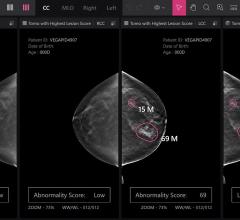
Despite decades of progress in breast imaging, one challenge continues to test even the most skilled radiologists ...
October 24, 2025 
Technology
Heart Imaging Technologies released the WebPAX cardiac echo reporting module.
September 16, 2013 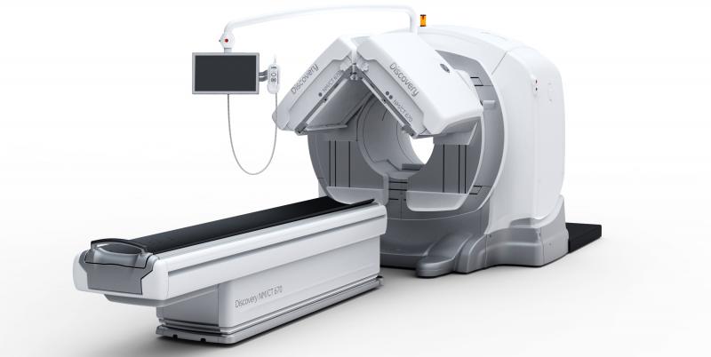
Feature | Dave Fornell
Diagnostic imaging is generally considered safe and noninvasive, so it is extremely unusual for a patient to die from injuries received from a scanner. However, this was the case in early June when a patient was killed because a portion of a SPECT/CT scanner fell during the scan at the James J. Peters VA Medical Center in the Bronx, N.Y.
September 13, 2013 Feature | Dave Fornell
The use of 3-D echo can help improve the accuracy and reproducibility of cardiac quantification. The technology has the advantage of removing the inter-operator variability by imaging whole volume datasets of the heart, so specific images or organ views can be extracted and reconstructed in any position, similar to CT or MRI datasets. Also, because a volumetric dataset is captured, exam times can be shortened, instead of spending time trying to get just the right angle for a 2-D slice view. Cardiac quantification can also be improved by measuring the entire heart or ventricle, rather than just slices of it. New software also automates this quantification.
September 13, 2013 Sponsored Content
Sponsored Content
Videos | Radiology Business
Bayer Radiology’s Barbara Ruhland and Thom Kinst discuss how radiology departments can address the many different ...
October 09, 2025 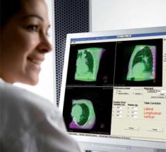
News
Integrated with Versa HD linear accelerator, Elekta has introduced new intra-fraction imaging capabilities within its X-ray Volume Imaging (XVI) system. During treatment, XVI now provides the tools to monitor and manage internal motion, supporting the clinician's efforts to increase therapeutic doses delivered to the tumor while reducing healthy tissue exposure.
September 13, 2013 
Feature
The Institute of Medicine (IOM) of the National Academies issued its report, “Delivering High-Quality Cancer Care: Charting a News Course for a System in Crisis.” The 315-page report, produced by a 17-member committee of cancer care leaders, concludes that the nation’s “cancer care delivery system is in crisis. Care is not patient-centered, many patients do not receive palliative care to manage their symptoms and side effects from treatment, and decisions about care often are not based on the latest scientific evidence.” In addition, the report details a six-part framework for improving the quality of cancer care, improving “the quality of life and outcomes for people facing a cancer diagnosis.”
September 13, 2013 
News
At RSNA 2013, Carestream’s new Vue PACS (picture archive and communication system) Reporting (shown as works-in-progress) is designed to enable insertion of key images and quantitative comparisons such as vessel analysis and measurements from modalities into radiology reports, which is designed to aid clinicians in treatment planning.
September 12, 2013 Sponsored Content
Case Study | PACS
eHealth Saskatchewan plays a vital role in providing IT services to patients, health care providers, and partners such ...
February 03, 2025 
Technology
The QA modules of Standard Imaging Inc. PIPSpro feature a new, simplified user-interface for TG-142 testing. Since the ...
September 12, 2013 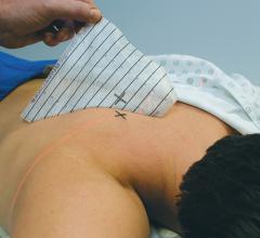
Technology
A light, medical grade, all-over adhesive ensures Beekley Medical’s new Mark-Through GuideLines stay in place without buckling or lifting during computed tomography (CT)-guided biopsies. Marks can be made anywhere on grid. The ink instantly transfers through to clearly marked entry sites on patient’s skin. Evenly spaced, lead-free radiopaque lines provide a distinct artifact-free image further enhancing the accuracy of needle placement and mark correlation.
September 11, 2013 News
At RSNA 2013, Toshiba will introduce a new single-panel Radrex-i wireless digital radiographic (DR) system X-ray system as a works-in-progress. The single panel wireless solution lowers cost of ownership while enabling more out-of-Bucky work and increasing workflow with one of the lightest detectors on the market.
September 11, 2013 
News
UltraSPECT announced that MaineHealth, a family of hospitals and medical centers throughout Maine, has selected the UltraSPECT Xpress.Cardiac and Xpress3.Cardiac solutions as part of the organization’s strategy for reducing nuclear medicine (NM) dose and complying with the American Society of Nuclear Cardiology (ASNC) final guidelines effective Jan. 1, 2014. Maine Medical Partners (MMP) – MaineHealth Cardiology in South Portland is one of ten facilities within MaineHealth that is installing the product, with others scheduled to continue roll out before the end of 2013.
September 11, 2013 News
Cancer patients from all over the world now have a new opportunity to access proton therapy today – at the Proton Therapy Center Czech in Prague. It is only the fifth such center in Europe and currently the most well equipped centre in the world. The facility is attracting child and adult cancer patients from around the world seeking advanced cancer care with few treatment-related side effects.
September 11, 2013 News
In patients with atrial fibrillation, delayed enhancement magnetic resonance imaging (DE-MRI) performed before ablative treatment can stage the degree of damaged heart tissue (atrial fibrosis) and help predict whether treatment will be successful or not, according to results of Delayed Enhancement — MRI determinant of successful Catheter Ablation of Atrial Fibrillation (DECAAF) trial.
September 10, 2013 
News
At RSNA 2013, Sony will be showing a works-in-progress medical recorder, the HVO-500MD (without DVD optical drive) and the HVO-550MD (with DVD optical drive). These models are designed specifically for medical recording.
September 10, 2013 
Technology
Toshiba is introducing a new Aquilion One platform at RSNA 2013. The new Aquilion One family allows providers to more precisely choose the system that fits their needs today while giving them an upgrade path for the future.
September 10, 2013 © Copyright Wainscot Media. All Rights Reserved.
Subscribe Now