July 10, 2025 — Fujifilm Healthcare Americas Corp. has launched several advanced automated functions for its FDR Visionary Suite digital radiography room. Optimized to support high-volume imaging in hospital radiology departments and imaging centers, the automation features are designed to enhance
-

-

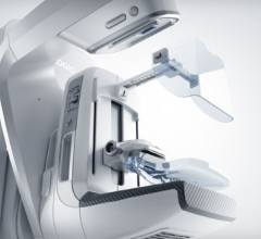
June 26, 2025 — Siemens Healthineers has received Food and Drug Administration clearance for the Magnetom Flow.Ace, its first 1.5 tesla (T) platform for magnetic resonance (MR) imaging with a closed helium circuit and no quench pipe, significantly reducing reliance on helium. The system, which has a
-
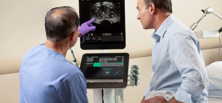
June 16, 23025 —GE HealthCare has launched the bkActiv S series, representing the next era of intraoperative ultrasound. The bkActiv S series is part of the bkPortfolio family of Active Imaging systems, designed to provide interventional guidance during urology, colorectal and pelvic floor
-

June 17, 2025 — Royal Philips has announced the global launch of the Flash Ultrasound System 5100 POC — a new point-of-care (POC) ultrasound system engineered for the fast-moving needs of anesthesia, critical care, emergency medicine and musculoskeletal imaging. Built on Philips’ established
-
![CEDM[1] copy](/sites/default/files/styles/slider/public/CEDM%5B1%5D%20copy.png?itok=XK5AvT8Q)
In breast cancer detection, speed and accuracy are more than clinical goals – they can significantly increase chances for cure and impact treatment paths and outcomes. Too often, women face barriers to advanced imaging due to cost, access, or availability. As healthcare systems seek solutions that

News | Radiology Imaging
July 15, 2025 — Radiology Partners (RP), a provider of technology-enabled radiology services, has announced the launch ...
 July 15, 2025
News | Radiology Imaging
The American Society of Radiologic Technologists (ASRT), in Albuquerque, N.M., was recently recognized by the ...
July 15, 2025 News | Ultrasound Imaging
July 14, 2025 — Patients with liver diseases will have expanded access to advanced ultrasound imaging and transplant ...
July 14, 2025 Sponsored Content
Case Study | PACS
eHealth Saskatchewan plays a vital role in providing IT services to patients, health care providers, and partners such ...
February 03, 2025 
News | Radiology Imaging
July 10, 2025 — Bracco Imaging was recently awarded a Platinum Medal by EcoVadis, one of the world's most trusted ...
July 11, 2025 
News | Digital Radiography (DR)
July 10, 2025 — Fujifilm Healthcare Americas Corp. has launched several advanced automated functions for its FDR ...
July 10, 2025 Sponsored Content
News | Artificial Intelligence | June 21, 2024
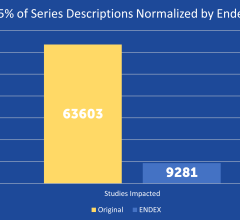
SPONSORED CONTENT — EnsightTM 2.0 is the newest version of Enlitic’s data standardization software framework. Ensight is ...

News | Lung Imaging
June 9, 2025 — bioAffinity Technologies, Inc., a biotechnology company addressing the need for noninvasive, accurate ...
July 10, 2025 Sponsored Content
Feature | Breast Imaging
While most women understand the importance of health screenings, an estimated 72 million have missed or postponed a ...
December 03, 2024 
News | Prostate Cancer
FDA Grants Breakthrough Device Designation for Artera's AI-Powered Software for Prostate Cancer Care
July 9, 2025 — Artera, the developer of multimodal artificial intelligence (MMAI)-based prognostic and predictive cancer ...
July 09, 2025 
News | ASTRO
July 9, 2025 — The American Society for Radiation Oncology (ASTRO) selected 43 members to receive the ASTRO Fellow ...
July 09, 2025 
News | Breast Imaging
July 8, 2025 — QT Imaging Holdings, has appointed Elaine Iuanow, MD, as chief medical officer (CMO) and Kim Du as senior ...
July 09, 2025 Sponsored Content
Fujifilm’s APERTO Lucent is a 0.4T mid-field, open MRI system addressing today’s capability and image quality needs ...
September 25, 2024 
News | Artificial Intelligence
July 2, 2025 — Lunit, a provider of AI for cancer diagnostics and therapeutics, has announced a collaboration with ...
July 08, 2025 
News | Breast Imaging
July 7, 2025 — SimonMed Imaging, one of the largest outpatient medical imaging providers in the United States, has ...
July 08, 2025 
News | FDA
July 8, 2025 — Mendaera, Inc., a healthcare technology company focused on developing robotics that can be deployed ...
July 08, 2025 Sponsored Content

News | Computed Tomography (CT)
SPONSORED CONTENT — Fujifilm’s latest CT technology brings exceptional image quality to a compact and user- and patient ...
August 06, 2024 
News | Computed Tomography (CT)
July 01, 2025 — NANO-X Imaging Ltd. recently announced a clinical and educational collaboration with Keiser University ...
July 03, 2025 
July 2, 2025 — Philips has received FDA 510(k) clearance for SmartSpeed Precise[1] MR’s latest deep learning ...
July 03, 2025 
Feature
A new study published in JCO Clinical Cancer Informatics tackles a critical challenge in cancer diagnostics: ensuring ...
July 01, 2025 
News | Ultrasound Imaging
July 1, 2025 — UPDATE: The final paper is now available at: JMIR AI - ChatGPT-4–Driven Liver Ultrasound Radiomics ...
July 01, 2025 
News | Ultrasound Imaging
June 26, 2025 — FUJIFILM VisualSonics Inc., a provider of ultra-high frequency ultrasound and photoacoustic imaging ...
June 27, 2025 
News | Radiology Imaging
June 18, 2025 — Probo Medical, a provider of medical imaging equipment, service, and support, has announced a strategic ...
June 26, 2025 
June 26, 2025 — Siemens Healthineers has received Food and Drug Administration clearance for the Magnetom Flow.Ace, its ...
June 26, 2025 
News | Prostate Cancer
June 26, 2025 – Quibim, a global provider of quantitative medical imaging solutions, has launched AI-QUAL, a new feature ...
June 26, 2025