With an increased availability of effective imaging techniques, peripheral vascular disease diagnosis and treatment is continuously advancing. Procedures no longer rely solely on the seemingly archaic practice of using simple 2-D fluoroscopic images in the vascular lab to administer treatment. Interventional radiologists now have much more at their disposal. Some of the most notable areas of recent development have been in the interventional radiologists’ abilities to acquire 3-D at a digital flat-panel level of image quality in the vascular lab during the procedures.


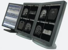
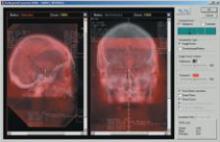
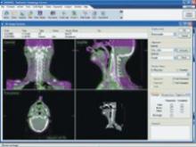
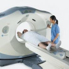
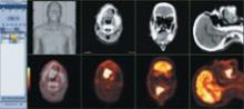
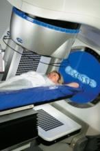
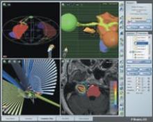
It’s all in the image – well, at least 90 percent anyway. Many radiologists will contend that optimum image quality and accurate diagnosis are directly related. Along this line of thought, healthcare has invested heavily in imaging equipment and software with higher grayscale contrast and more megapixels to render a sharper image. The use of multislice CT scanners has exploded in the last few years in an effort to capture greater anatomical detail. All of this has been geared toward enhancing clinical images to improve accuracy in diagnosis.


Despite government initiatives and encouragement for the U.S. healthcare industry to build a national electronic health record (EHR), the competitive nature of the U.S. healthcare system creates many obstacles, such as noninteroperability and software implementation costs, which have hindered progress.


Integrating disparate systems poses serious challenges to healthcare as information silos separate clinical information flow amongst radiology, oncology, cardiology and more.
Over the years, radiology has led the way in breaking down these silos and linking them across the enterprise. Now, radiology is again positioned to play the lead role in connecting imaging data sets and clinical information networks into a single national electronic health record (EHR).


The U.S. has made major strides in the implementation of healthcare information technology (IT) in the last several years, and while much work remains to be done, the future of healthcare IT in the country looks promising.


Since the 1990s, Americans’ risk of dying from cancer has continued to drop, according to a new report from the nation’s leading cancer organizations.* While lifestyle improvements and screening tools do affect cancer rates, the decline in cancer death rates can partly be attributed to advanced imaging and treatment technologies.


For years, intensity modulated radiation therapy (IMRT) has assisted doctors in contouring radiation beams around anatomical structures to target tumors. As tumors or organs shift, however, the beam can fall off target and damage surrounding healthy tissue. With image-guided radiation therapy (IGRT), which allows physicians to scan and locate the tumor while the patient is in the treatment position, the volume of healthy tissue exposed to radiation during treatment has shrunk from half an inch or more


Siemens Medical Solutions introduced syngo WebSpace, a client-server computed tomography (CT) workplace solution, at the Stanford Radiology 8th Annual International Symposium on Multidetector-Row CT.

Philips has released Model Based Segmentation (MBS) software for IGRT workflow enhancement. The software will be an upgrade on the Pinnacle3 Radiation Therapy Planning system.

MVision, from Siemens Medical Solutions, is a volumetric inline target imaging solution and the natural next step in Image-Guided Radiation Therapy (IGRT). Designed to work with Siemens’ linear accelerators, the system is the first commercial implementation of cone-beam technology utilizing a standard radiotherapy treatment beam. MVision makes it possible for the megavoltage (MV) source used for treatment to also create a 3-D image of the patient, enabling clinicians to “see inside” the patient at the most appropriate moment.

Subscribe Now