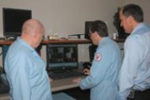
The Precess MRI Patient Monitoring System features wireless ECG and SPO2. The wireless ECG and SPO2 technologies allow for shorter cable lengths, which can increase patient safety and throughput.

Medical Imaging Resources is going places — to growing hospitals in need of faster MRI exam turnaround, to busy medical centers that benefit from back-up CT scanners, to facilities awaiting new equipment shipments and to the forefront of today’s rapidly accelerating mobile imaging scene.
“We started with a single mobile unit 15 years ago,” said John Vartanian, president of the Ann Arbor, MI-based mobile service.

Increasingly, U.S. community cancer centers are among the diverse clinical environments pioneering the use of Elekta Synergy systems, the leading commercial system for treating cancer patients using Intensity Modulated Radiation Therapy (IMRT) and Image-Guided Radiation Therapy (IGRT).

In March of 2006, clinicians at the MIMA Cancer Center in Melbourne, FL, began treating lung cancer with stereotactic body radiosurgery, delivered using the Trilogy medical linear accelerator from Varian Medical Systems, configured with the On-Board Imager device for image-guided radiotherapy and the RPM respiratory gating system for managing tumor motion.

Increasing System Speed
Exempla Healthcare has a legacy in Denver, CO having provided 130 years of comprehensive medical services. In pursuing their goal of becoming "Best in the Nation", they strive to continually improve every aspect affecting patient care. One area their IT department has focused on is increasing the radiology image transfer speeds throughout their PACS enterprise, so critical information reaches the radiologist and other healthcare providers faster.

CentraState Medical Center, a 270-bed acute care facility, belongs to CentraState Healthcare System, a nonprofit community health organization that has served Freehold, NJ since 1971.
CentraState’s Radiation Oncology Center opened its doors in April 2002 and, in 2006, was reaccredited by the American College of Radiologists (ACR) for three years. The program was also selected as one of 60 clinical trial sites in the nation by the Cancer Trials Support Unit of the NCI and The Cancer Institute of New Jersey’s Oncology Group.

With the introduction of recently FDA cleared MVision megavoltage cone-beam imaging package by Siemens Medical, Imaging Technology News (ITN) spoke with Andreas Schlatter, director of Marketing Development for Siemens Medical Solutions Oncology Care Systems, about the highlights of the first commercial implementation of cone-beam technology utilizing a standard radiotherapy treatment beam.


The name of the game in radiosurgery and radiotherapy is delivering the highest possible dose to the target volume and sparing the surrounding normal tissue and the structures. That requires immobilizing the target and drawing up a good plan, according to E. Ishmael Parsai, Ph.D., chief of Medical Physics, Medical College of Ohio, Toledo. The key to creating an individualized plan for accurate dose delivery for each patient goes beyond sophisticated software — it takes teamwork.


With an increased availability of effective imaging techniques, peripheral vascular disease diagnosis and treatment is continuously advancing. Procedures no longer rely solely on the seemingly archaic practice of using simple 2-D fluoroscopic images in the vascular lab to administer treatment. Interventional radiologists now have much more at their disposal. Some of the most notable areas of recent development have been in the interventional radiologists’ abilities to acquire 3-D at a digital flat-panel level of image quality in the vascular lab during the procedures.


It’s all in the image – well, at least 90 percent anyway. Many radiologists will contend that optimum image quality and accurate diagnosis are directly related. Along this line of thought, healthcare has invested heavily in imaging equipment and software with higher grayscale contrast and more megapixels to render a sharper image. The use of multislice CT scanners has exploded in the last few years in an effort to capture greater anatomical detail. All of this has been geared toward enhancing clinical images to improve accuracy in diagnosis.


Subscribe Now