For better or worse, healthcare reform has arrived, and with it comes a contentious debate about the certification of electronic medical records (EMR) systems for meaningful use. Managing large medical image files has long been a challenge even within radiology, yet diagnostic data would seem essential to a physician’s treatment of disease.


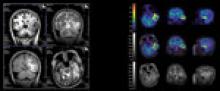
In scientific research, sometimes simply connecting two points with a line is enough to paint the big picture. When researchers first identified the biomarker amyloid beta-protein for Alzheimer’s disease by using positron emission tomography (PET) imaging, they gained a new perspective on how to treat the disease.


Editor's Note: This article is an introduction to the ultrasound scanning systems comparison chart that ran in the September 2010 issue of Imaging Technology News. The chart can be found under the comparison chart tab at the top of the page.


The applications for ultrasound in medicine have been quite varied. Back in the 1940s, physical therapists used its thermal energy to reduce inflammation, and physicists used high-intensity ultrasound as a surgical tool to treat brain disorders.


Despite the continued attacks by policymakers regarding imaging and its impact on healthcare costs, the diagnostic imaging industry is positioned to take on these challenges to not only survive, but also to thrive in the healthcare reform implementation era.


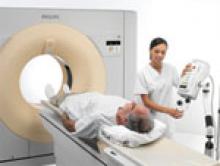
Advancements in newer 64-slice computed tomography (CT) and the introduction of 256- and 320-slice systems are helping to significantly reduce patient exposure to ionizing radiation.


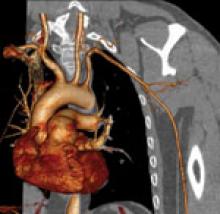
In this era of financial cost cutting, questions are raised over the true need for 256- or 320-slice computed tomography (CT) systems for cardiac imaging. There are many radiologists and cardiologists who argue a 64-slice CT system is good enough to make a cardiac diagnosis without spending twice as much for the higher-slice systems.


Biopsies were once the domain of surgeons, but with the introduction of image-guided needle biopsies in 1992, radiologists have increasingly taken over that diagnostic role.


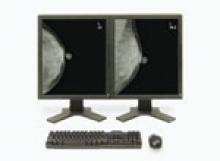
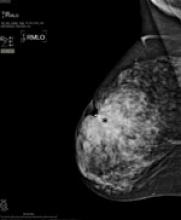
Mammography and breast magnetic resonance imaging (MRI) are tried-and-true methods for spotting and preventing the spread of breast cancer and other abnormalities. Mammography alone detects up to 90 percent of breast cancers in women over 50, according to the U.S. Food and Drug Administration (FDA).


Information-based decision-making is critical to physicians in delivering quality care and ensuring efficiency in today’s health care systems. Patient data must be available and used for effective decision-making. Yet, because it exists in various systems across numerous healthcare providers, this information is not always readily available to clinicians.


Subscribe Now