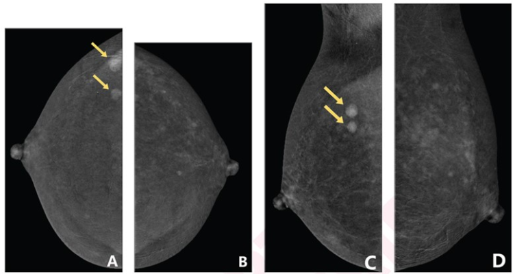
48-year-old woman who underwent CEM to evaluate two masses (arrows) in right breast. CEM obtained in following order: (A) craniocaudal (CC) view of right breast; (B) CC view of left breast; (C) mediolateral oblique (MLO) view of right breast; and (D) MLO view of left breast. Degree of background parenchymal enhancement (BPE) increases over time after contrast agent injection. (A) CC view of right breast shows mild BPE, whereas subsequently obtained (B) CC view of left breast shows moderate BPE. Based on criteria 1 reflecting only first obtained CC view, degree of BPE is classified as mild; based on criteria 2 reflecting both obtained CC views, degree of BPE is classified as moderate. Histopathologic assessment from subsequent biopsy demonstrated both masses to be invasive ductal carcinoma.
January 27, 2023 — According to an accepted manuscript published in ARRS’ American Journal of Roentgenology (AJR), both menstrual status and menstrual cycle time are associated with the degree of early background parenchymal enhancement (BPE) on contrast-enhanced mammography (CEM), which could influence CEM interpretations.
“Clinical factors, including history of breast cancer or breast cancer treatment, breast density, menstrual status, and time of menstrual cycle, are associated with degree of early BPE on CEM,” wrote corresponding author Yajia Gu from the department of radiology at Fudan University Shanghai Cancer Center in China. “In premenopausal patients, degree of BPE is lowest on days 8–14 of the menstrual cycle.”
This AJR accepted manuscript included 207 patients (median age, 46 years) who underwent CEM between April 2020 and September 2021. Two radiologists independently assessed BPE degree on CEM (minimal, mild, moderate, or marked) based on two criteria: using the first of four obtained views; using the first two of four obtained views. Reviewing electronic medical records for clinical factors, the radiologist pair reached consensus for breast density via CEM.
Ultimately, degree of early BPE on CEM was negatively associated with age, history of breast cancer, chemotherapy or radiation therapy, as well as perimenopausal and postmenopausal status. Meanwhile, early BPE degree on this modality was positively associated with dense breasts and premenopausal status with irregular cycles.
Acknowledging their retrospective study’s small sample size, given the potential impact of BPE on diagnostic performance, “the findings have implications for CEM scheduling and interpretation,” the authors of this AJR accepted manuscript added.
For more information: www.arrs.org
Related Breast Imaging Content:
Single vs. Multiple Architectural Distortion on Digital Breast Tomosynthesis
Today's Mammography Advancements
Digital Breast Tomosynthesis Spot Compression Clarifies Ambiguous Findings
AI DBT Impact on Mammography Post-breast Therapy
ImageCare Centers Unveils PINK Better Mammo Service Featuring Profound AI
Radiologist Fatigue, Experience Affect Breast Imaging Call Backs
Fewer Breast Cancer Cases Between Screening Rounds with 3-D Mammography
Study Finds Racial Disparities in Access to New Mammography Technology

 June 04, 2026


